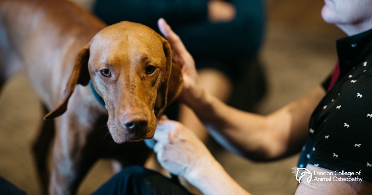
Osteopathy for Equine TMJ Dysfunction
Click to view
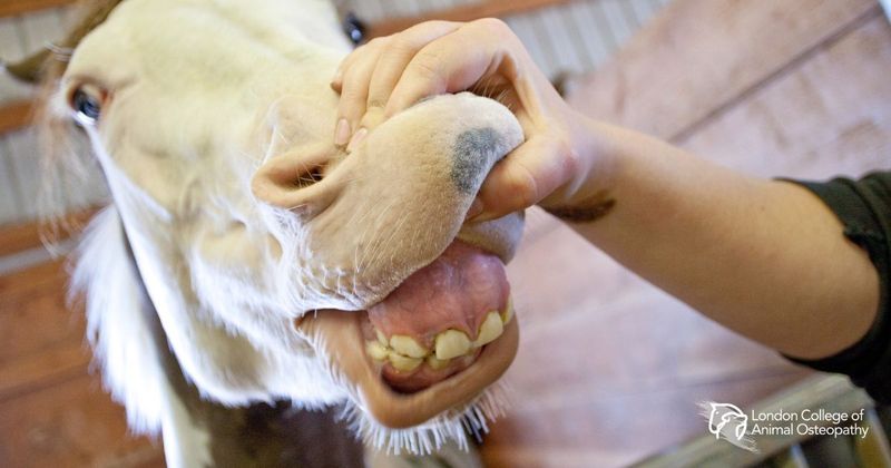
A horse grazes for up to sixteen hours a day under natural conditions. That means the temporomandibular joint — the TMJ — is one of the most continuously loaded structures in the equine body, yet it remains one of the least studied. While human TMJ dysfunction has attracted decades of clinical research, the equine equivalent sits in a diagnostic grey zone, where vague and overlapping symptoms make it difficult to distinguish a functional problem from outright pathology.
This thesis by Hanna Musakka sets out to map what is known about the equine TMJ, examine the range of dysfunctions that may arise, and assess whether osteopathic manipulative treatment — so well-established in human TMJ care — can be rationally applied to horses. The investigation draws on equine anatomy, veterinary literature, and a substantial body of human osteopathic research, building a case for cross-species application where direct equine studies remain scarce.
The anatomy alone is revealing. Equine TMJ fibrocartilage shows a unique zoning pattern suggesting higher regenerative capacity than its human counterpart — which may explain why clinical TMJ pathologies appear relatively rare in horses, or simply why they go undetected. The joint’s proximity to the hyoid apparatus, the guttural pouch, and multiple cranial nerves means that dysfunction here can cascade into problems that look nothing like a jaw complaint: headshaking, bit resistance, altered head carriage, even recurrent colic.
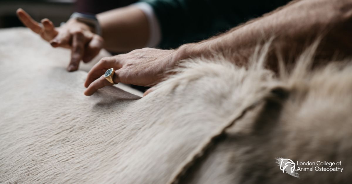
Musakka surveys the osteopathic literature on human TMJ treatment — myofascial release, muscle inhibition, balanced ligamentous tension, joint mobilisation — and makes a carefully reasoned argument for adapting these approaches to the horse. The cervicomandibular relationship proves particularly important: manipulation of the upper cervical spine in humans consistently improves mouth-opening range, pointing to a biomechanical link that almost certainly holds across species.
The thesis is candid about the limits of current knowledge. Without equine-specific clinical trials, the transfer of human treatment protocols requires both theoretical grounding and hands-on caution. Musakka, a licensed equine massage therapist turned osteopath, brings both — and shares her own clinical observations from treating horses presenting with hypertonic masticatory muscles and hyoid tension. The results, she notes, have been encouraging.