By Chris Bates M.Ost, DipAO, EEBW, BHSAI
One of the first things I get asked in my work as an Osteopath is “What is the difference between Osteopaths, Chiropractors, and Physiotherapists?” The fact of the matter is, many people have no idea what the difference is and why there are so many types of practitioners. I could be glib and say that variety is the spice of life, but there is so much to be gained by informing people of the aims, developments, and histories of the different practices. By giving our clientele the details around these distinct disciplines, they can make informed decisions about their animal’s care, and we will know that we are being called out to cases within our scope of practice.
Why Is It Important to Know the Difference Between Animal Therapists?
Distinct disciplines of therapy and wellness will approach the patient from different viewpoints. The goal of all approaches is one of achieving wellness, of course; however, the actual way they aim to achieve that can be vastly different.
Principles of practice could be described as the rules or laws that a discipline follows. Some therapies will have principles based upon years of trial and error, some will base theirs on repeatable evidence, and some will take more esoteric roots.
Similarities and Differences in Practice
When looking upon an Osteopath, a Chiropractor, and a Physiotherapist in practice, it may seem that there is little difference between them. This is because there are certain similarities in the way the animal is assessed and the way some physical methods of examination and treatment take place. The lens through which they each see the case in front of them, however, is different.
I must mention here that despite there being differences in the philosophy and viewpoint, this does not mean that they won’t come to the same or similar conclusions around causative factors of dysfunction and potential avenues towards health.
If an animal owner knows what each of the practitioner’s philosophies is (within reason), they can decide what approach they may need to take, and indeed, vets with this information can refer appropriately. In the case of an Osteopath being called out to attend an animal, and they feel the case is better served by a Physiotherapist, the referral process and loss of time only serve to increase the time before that animal can find relief.
The Importance of Proper Referral
As practitioners, there is a vital importance to knowing when a referral is needed. There are times when some practitioners may step outside their comfort zone, knowledge base, and scope of practice; this is, of course,e not professional or sometimes even legal. I have little doubt that they are only doing so to try and help, but the animal is the most important one, and admitting one’s limitations is the most helpful thing one can do.
Let’s face it, you don’t call a massage therapist when you’ve fallen off a roof and have a bone poking out of the skin, equally, you don’t go to A&E for a tight hamstring.
What is Animal Physiotherapy?
Physiotherapy, as it is often seen today,y is quite different from the methods used when it first became a part of conventional medicine. This is, in part, due to the technological advances made as modern Physiotherapy makes use of a variety of electrotherapy machines, mobility aids, and other medical equipment such as respiratory support.
The History of Physiotherapy
It could be argued that the origins of Physiotherapy go back beyond recorded history; there is evidence of modalities of touch and massage in historical and archaeological studies of cultures across the world. In ancient Greece, massage and manual therapy were used extensively by Hippocrates and Galenus. The ancient Romans used exercise and gymnastics to treat maladies and enhance the quality of life.
In these early times, however, the methods were simply part of general medicine and not a discipline in distinction from it. The early methods of Physiotherapeutic intervention were part of what physicians of the time used, and it was not so much the pursuit of a practitioner trained in only that.
Massage and exercise were the most notable aspects of Physiotherapy (as a medical approach), with some additional tools being used, such as wooden, metal,l and stone instruments to mobilise the tissues. Aspects of the eastern healing modalities, such as Traditional Chinese Medicine (TCM) or Indian Ayurveda, also used massage and advised on exercise; it is unknown historically how much influence these had on the European and Western development of Physiotherapy as a discipline.
The Foundation of Modern Physiotherapy
Massage was the initial definition of the practice, and in the UK, in 1894, the Society of Trained Masseuses was founded. This was the creation of four British Nurses: Lucy Robinson, Rosalind Paget, Elizabeth Manley, and Margaret Palmer. They had created the society to raise the profile of Massage and physical therapeutics inline with the medical field.
During the 20th century, the society gained a Royal Charter and eventually changed the name of the profession to “Physiotherapy” in 1944. In 1977, the profession gained professional autonomy, meaning that practitioners didn’t need patients to be referred by a physician. Degree courses were developed, and the continuous development of the profession led to statutory regulation by the Health Care Professions Council (HCPC).
Animal Physiotherapy Today
Physiotherapy also progressed very quickly in the animal world. Perhaps due to the higher prevalence of Physiotherapists being in recognised mainstream roles in Human practice, their profession moved into animal care very easily, and Vets had a clearer picture of what to expect of them.
Owners also seem to have a better grasp on what the Physiotherapist does, and this may be because they have experienced it themselves via mainstream healthcare. The initial organisations, courses,s and registers that developed in animal paraprofessional care were primarily Physiotherapy focussed.
Animal Physiotherapy courses are now available as:
- Undergraduate degrees (no previous Human Physiotherapy training needed)
- Postgraduate degrees/diplomas (some for Human trained Physiotherapists and some for those with relevant degrees)
- Diplomas (Ofqual registered and private)
If you’re considering becoming an Animal Osteopath, understanding the different pathways available is essential.
Physiotherapy Techniques and Methods
While many Animal Physiotherapists still utilise Massage, the primary focus in much of their training currently is exercise-based and the use of electrotherapies. There are many different movement and exercise approaches used, such as proprioceptive training, strength and conditioning, rehabilitation training, and sports-specific development.
Electrotherapies can include interventions like ultrasound, interferential, shockwave, TENS, Laser and many others. Most of the electrotherapies aim to stimulate muscle activation, increase localised perfusion, stimulate mitochondrial action, break down fibrous adhesions, or work on pain reduction. Some Physiotherapists may also use joint mobilisations and soft tissue massage.
While the Physiotherapists can work with all the same cases that other practitioners do, they have a particular strength in rehabilitation from injuries and optimisation of performance for active animals. Practitioners are highly skilled in owner education and helping people to understand their animals more deeply. This can involve providing tailored training and care techniques to enhance health and maintain well-being.
When to Refer to a Physiotherapist
I am an Osteopath, and so I can only comment on why I would refer to a Physiotherapist or use one myself. My referrals to them tend to be for focused rehabilitation and physical development after I have addressed a “lesion” or “configuration” from the Osteopathic perspective.
In cases of an animal’s conditions and symptoms arising from a weakness from poor exercise, handling, or environment, I will often seek a Physiotherapist to take the case to rectify these issues, as my skills are better put to use in the manual adjustment of the animal. When manual therapy is used in post-surgical rehabilitation, timing and patient selection are critical.
What is Animal Chiropractic?
While it is somewhat easier to understand the word “physiotherapy” (a therapy using physical means), the word “Chiropractic” does leave some people wondering. It is linking the root Greek words for “hand” and “done” to mean “Done by hand”. This reflects the heavy emphasis on the manual adjustments used.
The Origins of Chiropractic
Chiropractic was founded in America as a discipline (although D.D. Palmer was actually Canadian by birth) in 1895 by Daniel David Palmer (D.D. Palmer), who devised that bony misalignments or “Subluxations” could negatively influence the nervous system,m leading to a range of health conditions.
His hypothesis was that his manual adjustments of the body, predominantly the spine, could alleviate the conditions by returning the structure to its optimal position. It was in 1895 that Palmer performed an adjustment that reportedly restored a man’s hearing. In 1994, the Chiropractors Act was passed in the UK, providing themwith statutory regulation (for work on humans) and protecting the title.
Modern Chiropractic Practice
In Modern Chiropractic training, more of an evidence-based approach has been adopted, and although there are many methods and approaches that can trace back to early practice, most practitioners integrate a number of interventions such as exercise and lifestyle advice, and sometimes adjunctive treatments like Western acupuncture.
On the surface, it can seem that there is not much difference between Chiropractors and Osteopaths but the principles and often the methods used are different. Modern Chiropractors do use more in the way of soft tissue manipulation and other methods, but manipulation and thrust techniques are often still a mainstay of the profession globally.
The McTimoney Technique
There have been different schools of Chiropractic, with some,e such as the McTimoney School, teaching a more gentle approach using recoil manipulations. John McTimoney, who was an English Chiropractor created the McTimoney technique as a specialised and focused manipulation method that used the elastic recoil of tissues and was somewhat more gentle than some of the high velocity thrusts that were often synonymous with traditional Chiropractic.
It was the McTimoney technique that was pioneered in animal practice in 1954 in the UK.
Animal Chiropractic Training and Regulation
There are a number of organisations internationally that educate and govern Animal Chiropractic. In some countries, animal Chiropractic care is reserved only for Vets to perform; there are lobbies against this ruling.
Many animal Chiropractors are Human Chiropractors who have extended their training with postgraduate courses to be able to treat animals. However, there are now training pathways for Vets and non-therapists to train in animal Chiropractic techniques too. This has followed a similar course to the development of animal Physiotherapy education.
Most animal Chiropractic education focuses on the manual hands-on treatment of animals and some inclusion of exercise and rehabilitation; some practitioners will then add on additional interventions via short courses such as electrotherapies.
Chiropractic Philosophy and Principles
It is very common for Chiropractors to see similar cases as Osteopaths. Much of the time, it is a case of owner preference as to who they call out.
The actual principles of Chiropractic are vast and traditionally have much to do with what is described as “body intelligence” or “vitality,” which is seen as an energy of health that all creatures have. Initially,y the goal of Chiropractic was to allow for this vitality to act unobstructed and so allow the body to present health.
In Modern times, the training of Chiropractic has moved away from the somewhat esoteric concept of vitality and instead has embraced explanations of the methods’ effects on biological structures. A shared principle between Chiropractic and Osteopathy is that of the interrelationship between structure and function; this offers practitioners a way to see that perverted structure via movement, posture, compression, etc can alter the function.
There is a particular focus in Chiropractic principles around nerve function and the potential spinal interruptions of nerve function. This highlights how one could suggest that Chiropractors are spinal specialists, or at least spinal experts. In cases like trigeminal mediated headshaking in horses, understanding nerve function becomes particularly crucial.
My Experience Working with Chiropractors
The referrals between me and Chiropractors for animal patients (and humans) have mainly been when one of us can’t fit a patient in, which shows the similarities in practice. With continued professional development courses being developed that suit both Chiropractors and Osteopaths, the way practitioners develop once working is often very similar.
However, from my own personal and professional experience, and particularly in the animal industry, many Chiropractors rely heavily on the skeletal manipulations in their technique, such as the gentle McTimoney technique or the high velocity thrust techniques. It really comes down to finding the practitioner and methods that work for your animals and how your animals react to certain approaches.
What is Animal Osteopathy?
Founded in 1874 by Physician Andrew Taylor Still, Osteopathy was initially a medical profession to sidestep the sometimes damaging treatments of the time and to promote health by optimising the body’s own innate healing mechanisms. At the London College of Animal Osteopathy, we have courses on the history of Osteopathy so I won’t give away too much information here, but I will break down the objectives and approach.
Core Osteopathic Principles
Sharing the principle of structure and function being reciprocally interrelated with Chiropractic, Osteopathy also trusts in the natural ability to heal oneself with the right inputs and environment. Dr Still established the first Osteopathy school in Kirksville, Missouri, in 189,2 and since then, his students have spread Osteopathy globally.
The UK established statutory regulation for Osteopaths in 1993, and this provided confidence in the rigorous education and clinical experience needed to work as an Osteopath.
Osteopathy in the USA vs the UK
In the USA, however, Osteopathy became absorbed into the conventional medical field, and today, American Osteopaths are medical doctors who had some training in Osteopathic manual practice during their degree. The use of traditional manual Osteopathy in the USA is actually very low among the American Osteopaths (DOs). This has created some confusion across the world about Osteopathy’s identity and the scope of practitioners.
In the UK, Osteopathy has become more recognised by the National Health Service (NHS) and the mainstream medical community, with some Osteopaths now working in the NHS at various band levels. In the UK, however, Osteopathy maintains its traditional roots of manual intervention and upholds the principles of the early Osteopaths.
Osteopathic Philosophy and Holistic Approach
Osteopathic philosophy is one of holism and sees the person or animal as a triune of being: body, mind, and spirit. Over the years, Osteopathy has built a good evidence base and continues to commit to research.
The methods used by the Osteopath do include manipulation similar to Chiropractic; however, there are also a number of other techniques, such as soft tissue techniques, cranial Osteopathy, and visceral manipulation. Techniques like Equine CranioSacral Therapy exemplify the subtle approaches within osteopathy.
Traditionally,y the principle of circulatory quality was strong and would sit in the same importance as the Chiropractic importance of neural flow. The early writings in Osteopathy on the importance of fluid mechanics were also construed as the importance of uninterrupted communication, both afferent and efferent, whether that be fluid or neural.
Animal Osteopathy as a Growing Profession
In the animal industry, Osteopathy has grown but remains a smaller profession than the other two described above. This has been partly through the lack of choice in training courses and the regulations over the title “Osteopath” in the human therapy world.
The London College of Animal Osteopathy has created educational pathways internationally to spread the knowledge of Osteopathy to animals the world over, and we seek to grow the profession further. These programs offer comprehensive training, including the International Diploma in Animal Osteopathy.
Benefits of Osteopathic Treatment for Animals
Some animal owners like the gentle approaches Osteopaths can choose when treating, and many are amazed by the profound impacts they see. Taking the whole animal into consideration, Osteopaths are very well placed to find root causes and resolve chronic issues that may not respond to other methods. This is particularly evident when treating conditions like osteoarthritis in horses and dogs or managing hip dysplasia in dogs.
Osteopathy also fits very well into a maintenance routine, and many people will use Osteopaths to maintain performance in sporting animals. Canine athletes particularly benefit from osteopathic care for injury prevention and performance enhancement.
Working as a Multidisciplinary Team

All practitioners should know they are not alone and that working as a multidisciplinary team is the best way to ensure animal welfare. Within each of the above three professions, there will be many differences between the practitioners, and so you may find a Physiotherapist who works very much like an Osteopath or a Chiropractor who works very much with rehabilitation and Physiotherapeutic machines.
Importantly, all practitioners should only work within the scope of their training and experience.
How to Choose the Right Practitioner
A good start is to ask a potential practitioner about how and where they trained and what their practice ethos is. This way, you can also get an idea as to whether you “click” with them, as it is important that you trust them. A reputable practitioner will recognise their skill set, and in the event that they do not have the required knowledge or skills for a certain case, they will refer to a practitioner who does.
Some practitioners will specialise in a certain population of patients or a species; these people will sometimes develop specialised knowledge that makes them experts in their field and sought after as referral pathways. I personally specialise in Equine patients and have found that my client base has built up with the type of patients I enjoy.
This is not to say that those who are more generalist are any less useful. Generalist practitioners with a wide range of experience can often recognise things that more specialist people won’t.
Understanding When Each Therapy is Needed
Becoming educated on the principles and philosophies of the practitioner you wish to use can help you greatly. Remember that learning these disciplines takes years of dedication, and each has an important role to play.
Osteopathy and Chiropractic rely on the innate healing abilities of the body. If the animal’s vital reserve and healing capacity is overwhelmed by the injury or condition, then maybe an electrotherapy from a Physiotherapist will be required. If the animal is performing the exercises provided by the Physiotherapist but is building unevenly or reacting abnormally, perhaps there is a somatic dysfunction that an Osteopath or Chiropractor needs to address first.
The three disciplines here are also continually evolving with newer evidence, practitioners, and professional stakeholders work to drive innovation while maintaining the philosophical grounding that underpins them. Understanding aspects like the hidden role of fascia in animal movement can help practitioners across all disciplines improve their treatment approaches.
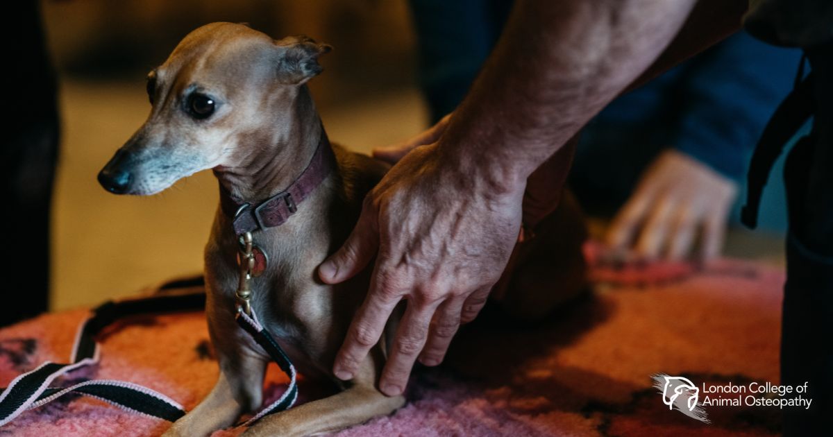
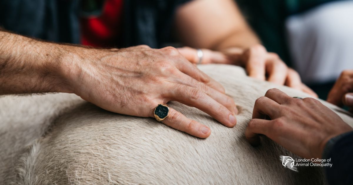
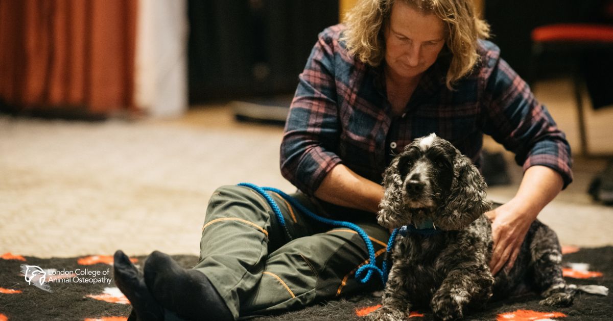
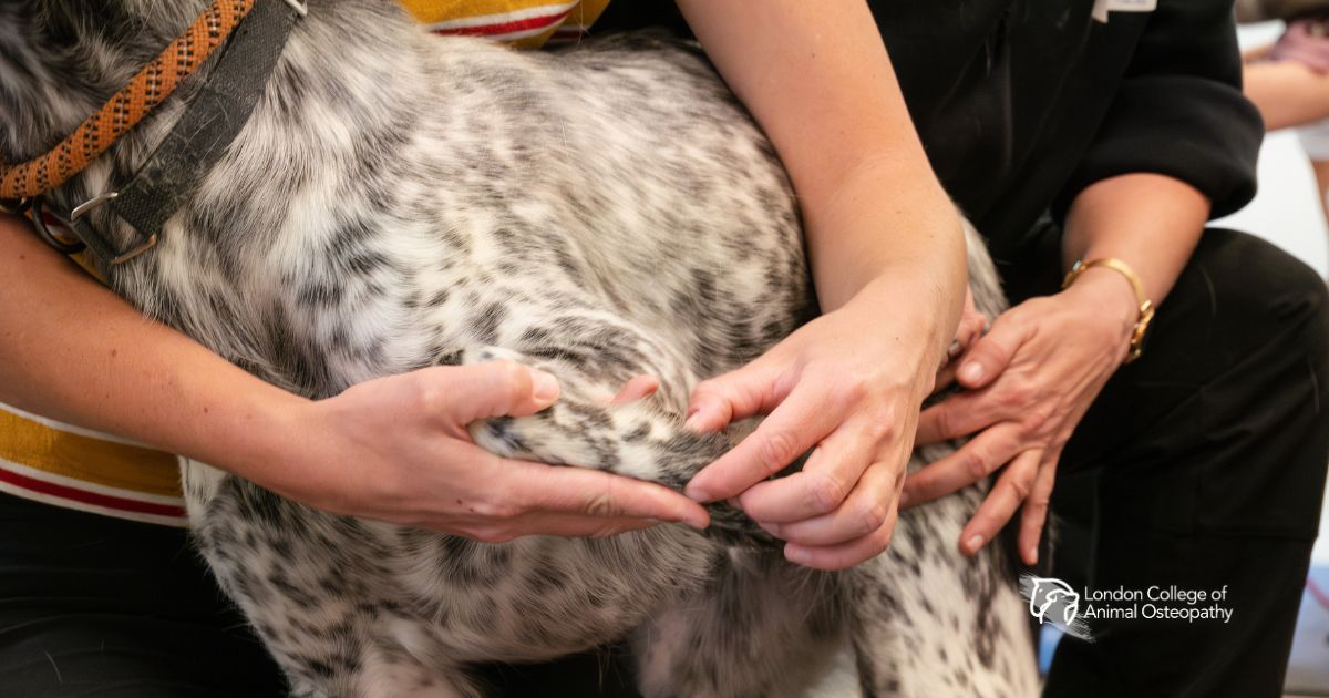
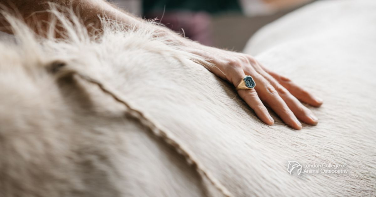
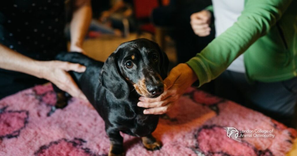
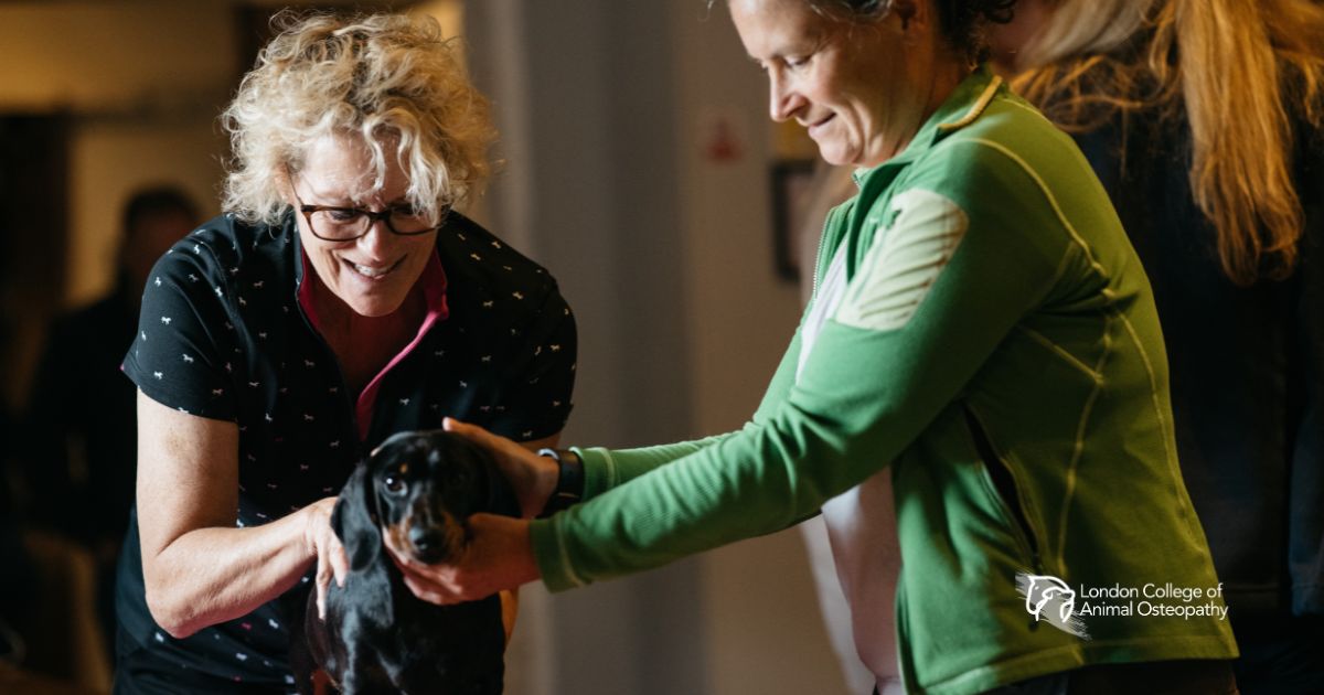 Manual osteopathic treatment is being applied to a dog, demonstrating hands-on assessment of musculoskeletal function
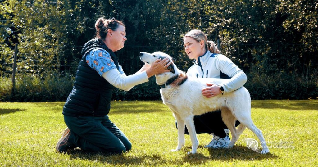
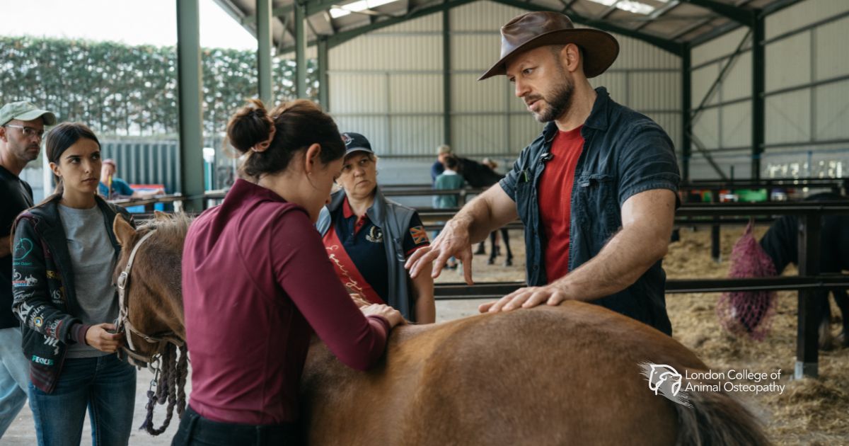
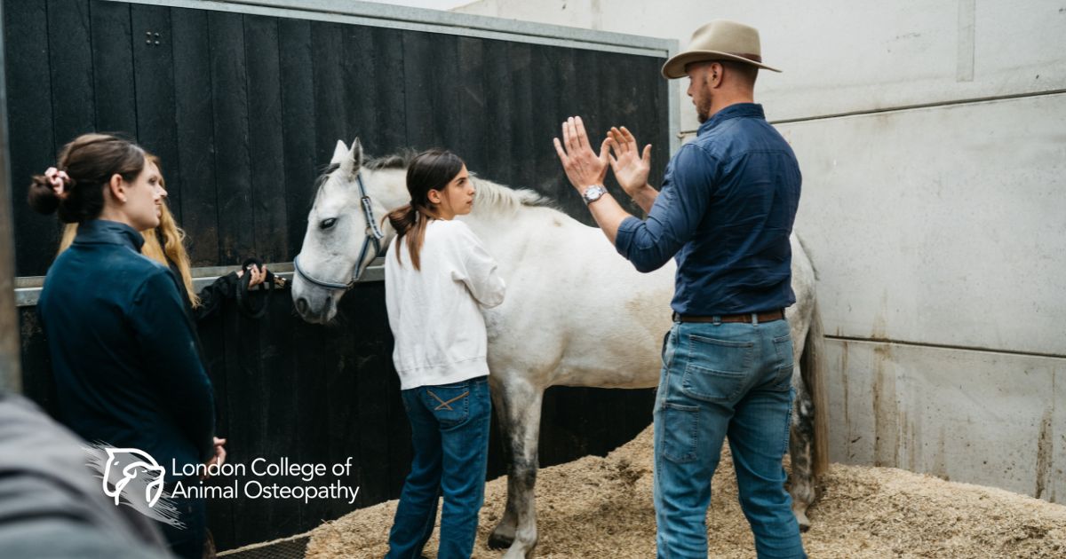
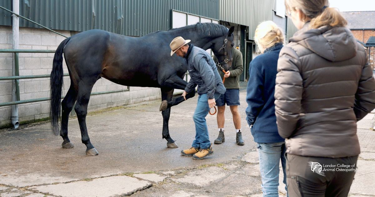
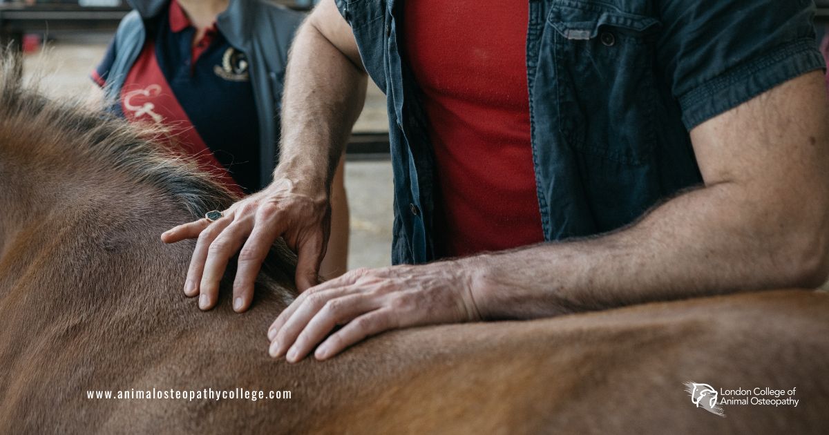
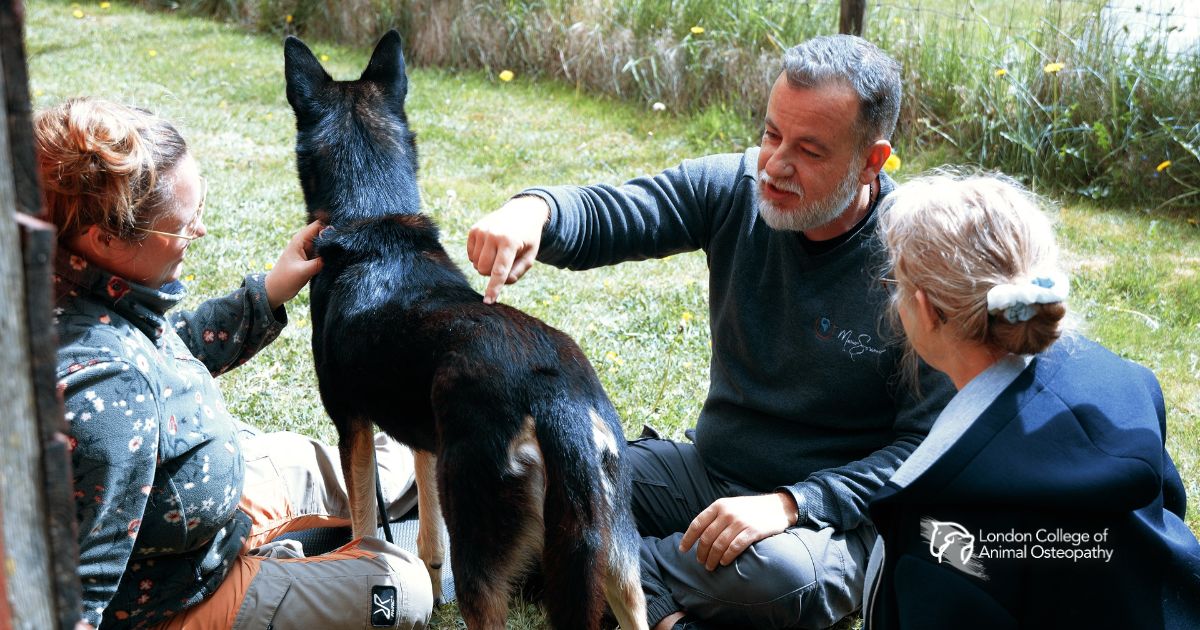 An animal osteopath working alongside a veterinarian, reflecting the collaborative approach central to professional animal osteopathy practice