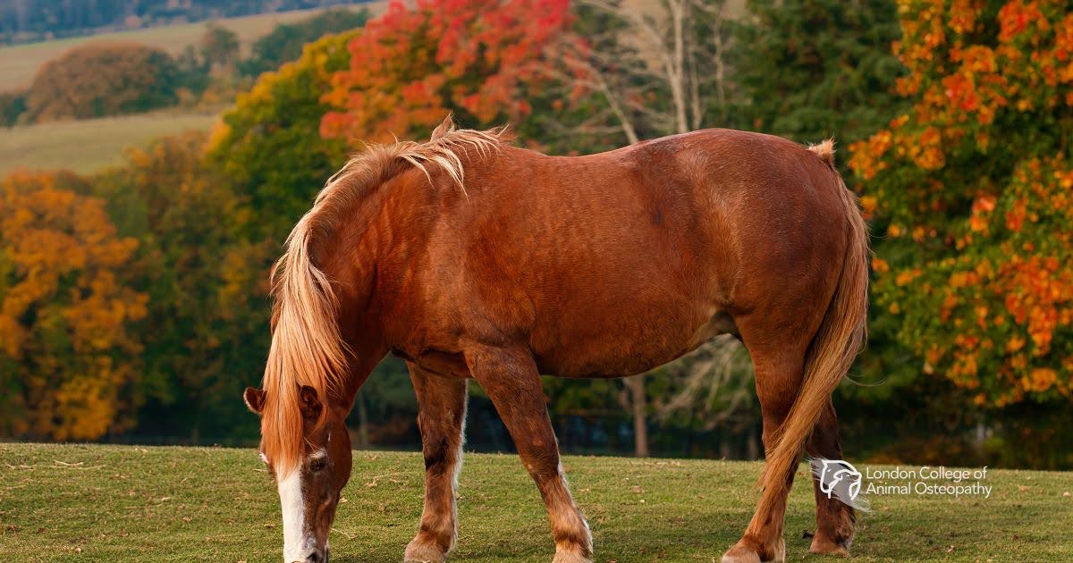
Autumn (Fall) Laminitis: Risks Often Overlooked

Autumn is sometimes thought of as a “safe” season for horses after the spring grass surge – but in many regions, it brings its own laminitis risks that are easily missed. Here we’ll take a look at why autumn can be hazardous, what usually causes laminitis at this time of year, modern veterinary treatments and hoof-management strategies, which practitioners should be involved, and a realistic, evidence-based take on when osteopathy can be appropriate during recovery.

Why autumn is risky (and which risks are often overlooked)

Non-structural carbohydrate (NSC) spikes in pasture
Grasses store carbohydrates (starch and fructans) in stems and roots. Warm, sunny days followed by cool nights – a common pattern in both spring and autumn – can raise fructan and sugar levels in pasture plants. High pasture NSC is a well-recognized trigger for pasture-associated laminitis in insulin-sensitive horses and ponies. Owners often focus on spring grass, but the same physiological plant responses make some autumn pastures risky too.

Management changes and “autumn flush” misconceptions
After hot, dry summers, a period of autumn rain and moderate temperatures can produce a sudden flush of leafy growth. Owners who relax grazing restrictions in autumn (thinking the risk is over) can inadvertently expose susceptible animals to high-NSC forage. Guidance is to monitor pasture NSC risk periods and manage turnout accordingly.

Endocrine causes remain the dominant factor
Across seasons, most laminitis cases in many populations are linked to insulin dysregulation (equine metabolic syndrome and PPID/Cushing’s). Autumn does not change that biology, but seasonal weight changes, feed changes, and grazing behaviour can interact with existing insulin dysregulation to precipitate an episode. In other words, autumn acts as a trigger on top of metabolic susceptibility.

Owners miss subtle early signs
Autumn laminitis is sometimes less dramatic than an acute overload case (eg, grain overload). Instead, owners may see mild foot soreness, changes in gait, short-stridedness on hard ground, or a “not quite right” attitude. Because the classic rocking-back laminitic stance or bounding digital pulses are not always present early, early cases may be missed or misinterpreted. Early veterinary assessment matters.

Common autumn precipitating mechanisms

  • Pasture-associated (forage high in fructans/NSC) – sudden exposure to high-NSC grass.
  • Insulin dysregulation (EMS) and PPID – chronic endocrine predisposition; autumn can be the season where management changes reveal the problem.
  • Obesity/weight gain through summer – autumn – more weight increases lamellar loading and risk.
  • Systemic disease/sepsis / grain overload/steroid administration – less season-specific but always important to consider.

Modern veterinary treatment: principles and evidence

Laminitis is a veterinary emergency. Once clinical signs appear, damage to the lamellae is already underway; treatment focuses on stopping progression, controlling pain, protecting the foot, and addressing the underlying cause. Key components:

Immediate veterinary assessment and triage
Rapid evaluation by a veterinarian determines severity, likely cause (endocrine vs inflammatory), and immediate steps (analgesia, stall rest, feeding management). Radiographs (podiatry views) are often taken early to document rotation/sinking and guide farriery.

Analgesia and anti-inflammatory management
NSAIDs (eg, phenylbutazone or flunixin) are commonly used to control pain; more severe pain may require multimodal analgesia under veterinary guidance. Acepromazine has historically been used in some protocols, but treatment must be individualized.

Dietary and management changes
For pasture-associated or endocrine cases: immediate removal from dangerous pasture, provision of low-NSC forage (soaked or tested hay, haylage with known low NSC), and strict weight management. For endocrine disease, long-term medical management (eg, pergolide for PPID) is indicated when diagnosed.

Foot protection and mechanical support (farriery)
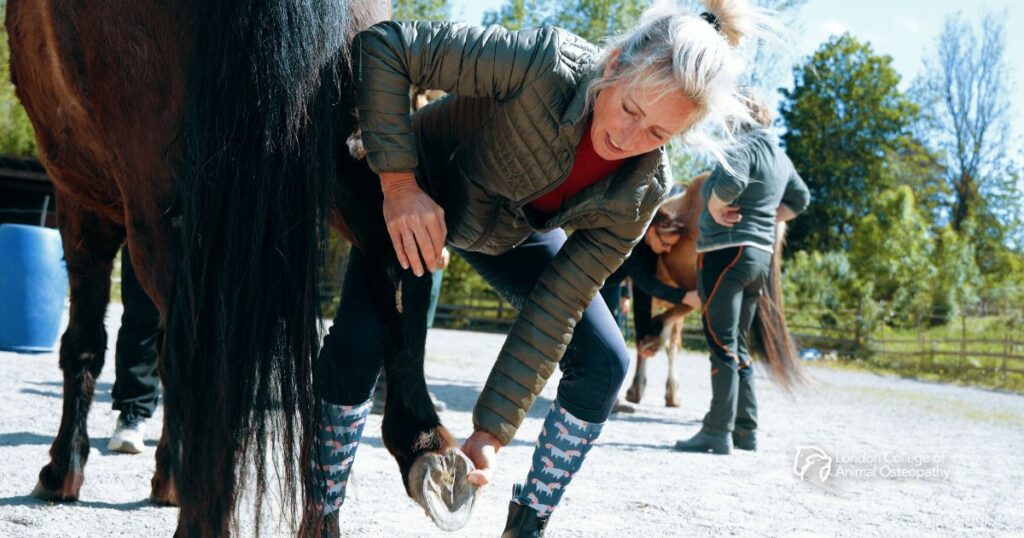
Farriery is central to limit mechanical trauma to the lamellae: deep, supportive bedding; frog/sole support (eg, impression material, pads); therapeutic trimming and specialized shoes (eg, heart bar, aluminium or foam supports) in subacute/chronic cases. Close vet–farrier cooperation is critical: radiographs guide trimming/shoeing plans and the timing of interventions.

Cryotherapy (digital hypothermia) — evolving, promising evidence
Experimental and clinical studies indicate that continuous, prolonged cooling of the distal limb (ice-water immersion or specialized cryotherapy devices) can markedly reduce lamellar injury if applied early. The strongest experimental evidence shows substantial protective effects when applied promptly; systematic reviews conclude the evidence is encouraging but call for further randomized clinical trials. Recent device developments (commercial cryotherapy systems and dry cryotherapy sleeves) improve feasibility in clinical practice. Cryotherapy is best directed by a veterinarian who understands timing, duration, and logistics.

Supportive medical care for systemic causes
If laminitis follows sepsis, endotoxaemia, or grain overload, treating the primary disease (fluids, antimicrobials where indicated, anti-endotoxin measures) is essential to reduce further lamellar injury.

Who should be involved — the full practitioner team

Effective laminitis care is multidisciplinary. The core team typically includes:

  • Veterinarian (primary clinician) — diagnosis, analgesia, metabolic testing (insulin, ACTH), medical management, radiographs, directing cryotherapy, and overall care plan.
  • Farrier/hoof care specialist — therapeutic trimming and shoeing, sole/frog support, regular podiatry follow-up. Their timing and technique must align with the veterinarian’s assessment and radiographic findings.
  • Veterinary nurse/technician — monitoring, bandage/ice boot application, medication administration, owner education.
  • Equine physiotherapist/rehabilitation specialist — once the horse is stable and weight-bearing, formal rehabilitation (controlled exercise, strengthening, balance work) can be implemented under veterinary guidance. Evidence is growing for structured rehab, but protocols must be individualized.
  • Nutritionist / veterinary nutrition advisor — for testing and planning low-NSC diets and long-term weight management.

Communication between these professionals and with the owner is repeatedly emphasised in the veterinary literature as a major determinant of outcome. Early engagement of a farrier and clear role-sharing with the veterinarian improve decision-making and owner compliance.

Osteopathy: when and when not to use it

Short answer: Osteopathic or other manual therapy can have a place as an adjunct in recovery, but only after the veterinary team has stabilised the laminitic foot and given clearance. There is limited direct, high-quality evidence for osteopathy specifically in laminitis, but there is veterinary literature supporting manual therapies for musculoskeletal dysfunction and gait issues more broadly. Use the following practical rules:

Do not use manual/osteopathic treatments in the acute, unstable phase.
While the foot is inflamed, painful, and at risk of ongoing lamellar failure, manipulation that encourages movement or weight-bearing changes (or that distracts from necessary stall rest and controlled support) is inappropriate. Acute management must be veterinary-led (analgesia, cryotherapy, mechanical support).

Once the horse is stable, weight-bearing, and cleared by the vet, osteopathy may help with secondary musculoskeletal issues.
After the laminitic event has been contained and mechanical hoof support established, many horses develop compensatory tension, altered posture, gait asymmetries, and back, sacro-iliac, or cervical dysfunction. Veterinary peer-reviewed articles show that spinal manipulation/osteopathic techniques can alter gait and address somatic dysfunction in horses. Manual therapies are a component of rehabilitation programs used by equine physiotherapists and osteopaths to restore functional symmetry, range of motion, and comfort. However, the evidence base is still small and mostly supportive rather than definitive.

Coordinate care: osteopaths must work under veterinary direction and in partnership with the farrier.
Any manual therapy plan should be integrated with ongoing farriery and veterinary monitoring (radiographs, lameness assessment). For example, changes in hoof mechanics after trimming or shoeing can alter the loading of limb segments; osteopathic work should reflect those mechanical realities. Communication and shared records are essential.

Be conservative with techniques that change limb loading or encourage early return to intense activity.
Rehabilitation after laminitis prioritises a gradual return to controlled exercise only after radiographic and clinical signs permit. Osteopathic treatment should support that goal — relieve compensatory muscle tension, encourage normal movement patterns in a carefully staged program — not shortcut it.

Practical autumn prevention checklist

  • Test hay/pasture NSC if possible, or follow local extension/veterinary advice about risky times. Restrict turnout when pastures are likely high in sugars (often late morning to afternoon; avoid flushes after rain following drought).
  • Identify at-risk animals (obese, cresty neck, history of laminitis, PPID/EMS) and implement stricter grazing control year-round.
  • Maintain a weight-management plan and consult a nutritionist/veterinarian for low-NSC forage options.
  • Build a veterinary–farrier relationship before problems occur; podiatry radiographs and a pre-agreed emergency plan save crucial time.

Bottom line

Autumn carries real laminitis hazards that are sometimes underestimated. The pathophysiology is usually endocrine (insulin dysregulation) with pasture or management changes acting as the trigger; pasture NSC/fructan spikes in cool nights/warmer days are a key mechanism. Prompt veterinary assessment, coordinated farriery, diet and weight control, and (where indicated) targeted treatments such as distal limb cryotherapy and medical management are the evidence-based pillars of care. Osteopathy and other manual therapies can be useful later in recovery to address compensatory musculoskeletal problems — but only as part of a coordinated, vet-led rehabilitation plan and not as a substitute for veterinary or farrier interventions.

Q1: Why is autumn considered a risky season for laminitis in horses?

Autumn pastures can be deceptively dangerous. During warm days followed by cool nights, grasses accumulate sugars and fructans (non-structural carbohydrates) that predispose insulin-dysregulated horses to laminitis. Owners sometimes assume the risk ends after spring, but the “autumn flush” of new grass and relaxed grazing restrictions can be just as hazardous. Horses with equine metabolic syndrome (EMS), PPID, or excess weight are particularly vulnerable.

Q2: What is the recommended modern treatment approach for a horse with acute laminitis?

Laminitis is a veterinary emergency. Current best practice includes immediate veterinary assessment, pain relief (usually NSAIDs), strict stall rest, and mechanical hoof support guided by radiographs. In suitable cases, cryotherapy (continuous digital hypothermia) can reduce lamellar injury if applied early. Long-term care involves weight and diet management (low-NSC forage), treatment of underlying endocrine disease (eg, pergolide for PPID), and close collaboration between vet and farrier for therapeutic trimming or shoeing.

Q3: Can osteopathy or manual therapy be used to help horses recovering from laminitis?

Not during the acute phase — when the foot is unstable and painful, only veterinary and farriery treatments are appropriate. However, once the horse is stable, weight-bearing, and cleared by a veterinarian, osteopathy may help address compensatory back or limb tension caused by altered posture and movement. It should always be integrated into a vet-led rehabilitation plan and coordinated with ongoing farriery adjustments.

Blog Post written by:

By Siun Griffin

Animal Physiotherapist and Community Manager at London College of Animal Osteopathy (LCAO).

Red Flags and Contraindications: When NOT to Use Certain Enrichment Activities

Enrichment is an essential component of canine well-being, but not all activities are appropriate for every dog. For the educated practitioner, recognising red flags and adapting plans is critical – not only to prevent injury but also to ensure that enrichment complements, rather than conflicts with, osteopathic care.

Why Contraindications Matter

Enrichment introduces variability in movement and environment, which is generally beneficial for tissue health and neural stimulation. However, in dogs with underlying musculoskeletal dysfunction, systemic illness, or pain-related behaviour, some activities can exacerbate problems. Overlooking these considerations risks compromising both physical integrity and recovery.

Key Red Flags Before Recommending Enrichment

  1. Acute Lameness or Sudden Postural Changes
    If a dog presents with new or worsening lameness, reluctance to bear weight, or asymmetrical posture, enrichment involving dynamic movement, such as balance work or tug games, should be avoided until a full assessment is performed. These signs may indicate joint pathology, muscle tear, or neurological involvement that requires veterinary evaluation.
  2. Signs of Pain During Activity
    Yawning, lip licking, tail tucking, or freezing mid-activity are subtle yet significant indicators of discomfort. Persisting despite these behaviours can lead to tissue overload, increased fascial tension, and compensatory strain. Activities should be paused and adapted to reduce intensity or eliminated altogether.
  3. Neurological Concerns
    Dogs showing ataxia, knuckling, or delayed proprioceptive responses should not engage in unstable surface work or agility-based enrichment. These activities require precise neuromuscular control and can increase the risk of falls or further neurological compromise.
  4. Excessive Fatigue or Stress
    Cognitive enrichment is beneficial, but prolonged problem-solving tasks can heighten stress in anxious dogs. Similarly, physically demanding enrichment in unfit or geriatric dogs can lead to muscular fatigue and joint overload. Monitoring duration and intensity is essential.

Activity-Specific Contraindications

  • Proprioceptive Equipment: Avoid in dogs with acute spinal issues, significant joint laxity (e.g., severe hip dysplasia), or post-surgical instability.
  • Tug Games: Contraindicated in dogs with cervical pain, temporomandibular dysfunction, or recent dental work.
  • Fetch or Jumping Activities: Should not be recommended for dogs with forelimb lameness, elbow dysplasia, or advanced osteoarthritis due to repetitive concussive loading.
  • Puzzle Toys: While generally safe, they may cause frustration in dogs with cognitive decline or those exhibiting compulsive behaviours – requiring careful observation.

Integrating Clinical Reasoning with Osteopathic Principles

The osteopathic approach emphasises the body’s ability to self-regulate and adapt – but only when conditions allow. Introducing enrichment during acute inflammatory stages or when pain inhibits normal motion can disrupt this process. Instead, enrichment should be phased in gradually, following tissue recovery and restoration of baseline mobility.

When to Refer Before Proceeding

Persistent pain, progressive neurological signs, systemic lethargy, or unexplained behavioural shifts warrant veterinary referral before implementing enrichment. Collaboration with the primary care veterinarian ensures that underlying pathology is addressed prior to introducing activities designed to enhance functional adaptability.

Final Thoughts

Enrichment is powerful, but not universally benign. Its effectiveness depends on timing, individualisation, and clinical judgment. By applying osteopathic principles alongside evidence-informed caution, practitioners can ensure that enrichment serves as a tool for healing and resilience rather than an unintended source of harm.

Practical Enrichment Strategies: Supporting Canine Musculoskeletal and Neurological Health

In the previous discussion, we examined enrichment as a concept that aligns closely with osteopathic principles of health maintenance and functional integrity. Now, let us move from theory to practice by exploring specific enrichment activities and how they influence the canine body on a structural and physiological level.

Designing Enrichment with Purpose

While all enrichment provides mental stimulation, activities should be chosen with intention, considering the dog’s physical condition, age, and behavioural tendencies. For practitioners with an osteopathic perspective, the objective is not only to engage the mind but to promote balanced movement, proprioceptive awareness, and tissue adaptability.

1. Scent-Based Work for Postural Adaptability
Example:
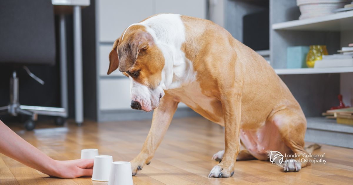
 Scatter feeding in varied terrain or structured scent trails.

Why It Matters: Searching for hidden food requires repeated cervical flexion, extension, and controlled shifts in weight-bearing as the dog tracks odour sources. These small postural changes improve cervical mobility and activate stabilising musculature in the thoracolumbar region. For dogs prone to stiffness, especially in the neck and forelimbs, this low-impact exercise provides dynamic movement without excessive load.

2. Proprioceptive Equipment for Joint Integrity
Example:
 Balance pads, wobble boards, or low cavaletti poles

Why It Matters: Engaging with unstable or varied surfaces stimulates joint mechanoreceptors, which in turn enhance neuromuscular coordination. From an osteopathic viewpoint, this is critical for maintaining the integrity of joints and preventing compensatory dysfunction. Cavaletti work encourages limb flexion and symmetrical stride length, supporting balanced gait patterns and reducing unilateral strain.

3. Textural and Surface Exploration for Fascia Health
Example:
 Walks incorporating sand, grass, gravel, or shallow water.

Why It Matters: Different surfaces require subtle adjustments in muscle tone and fascial tension, encouraging adaptability across kinetic chains. This variation helps maintain fascial glide and elasticity—an essential aspect of functional biomechanics. For osteopaths, such activities complement manual interventions by reinforcing tissue mobility through active movement.

4. Interactive Play for Spinal Mobility
Example:
 Controlled tug games or fetch on varied terrain.

Why It Matters: When executed with proper mechanics (e.g., avoiding vertical pulls during tug), these games provide dynamic spinal rotation and limb engagement. The oscillatory loading pattern during tug play can improve spinal flexibility and thoracic mobility, provided the movement is symmetrical and not excessive.

5. Problem-Solving Toys for Stress Regulation
Example:
 Puzzle feeders or DIY foraging boxes.

Why It Matters: Beyond cognitive stimulation, mental engagement reduces sympathetic nervous system overdrive, allowing the parasympathetic system to dominate – a state that promotes tissue healing and systemic balance. Osteopathic philosophy recognises the link between autonomic balance and structural health, making these activities valuable in a holistic care plan.

Integrating Enrichment into Osteopathic Care Plans

When advising clients, the practitioner should consider the timing and intensity of enrichment activities, particularly post-treatment. Low-impact sensory games may be introduced immediately after a session, whereas physically demanding exercises should be delayed until tissue recovery stabilises. Enrichment should not replace rest but should complement it, acting as a bridge between passive care and active functional restoration.

Final Thoughts

Enrichment is far more than a behavioural tool – it is an applied strategy for sustaining adaptability, reducing injury risk, and enhancing neuromuscular efficiency. When paired with osteopathic care, it becomes a potent method of maintaining health, preventing dysfunction, and honouring the principle that structure and function exist in a reciprocal relationship.

Stay tuned for part 3!

Equine Nutrition: Foundations for Health, Performance, and Longevity

Equine nutrition lies at the heart of any horse’s well-being, from a leisurely pasture companion to a champion athlete. As hindgut fermenters, horses digest fiber through a delicate, microbe-rich large intestine rather than through a multi-chambered stomach like ruminants. This fundamental difference means they thrive on frequent, steady intake of forage, yet remain exceptionally vulnerable to digestive upset when changes occur too rapidly.

Importance of Water

Water, often overlooked, is arguably the most critical nutrient. A horse’s body is roughly two-thirds water; even modest dehydration can be life-threatening. Clean, comfortable water intake supports everything from digestion to thermoregulation – simple, yet essential.

Horses require six essential nutrient categories: water, fats, carbohydrates, protein, vitamins, and minerals. Carbohydrates, especially non-structural ones, supply quick energy, while fiber is fermented in the hindgut into volatile fatty acids, offering sustained energy and supporting gut integrity. Protein supports growth and repair, while fats provide a calorically dense energy source, but must be introduced slowly, given horses’ limited capacity to digest fats efficiently.

Don’t Overlook Vitamins & Minerals

Vitamins and minerals also warrant attention. While quality forage often supplies most needs, imbalances may arise when diets lean heavily on grain, include low-grade hay, or the horse is under physical or environmental stress.

Equine Health & Wellbeing

Contemporary insight highlights how targeted nutritional adjustments can enhance coat condition, metabolic balance, musculoskeletal recovery, and overall vitality. For instance, omega-3 rich oils from sources like flax not only nourish the coat but also modulate inflammatory processes. Likewise, emerging work into gut microbiome support adds promising layers to our nutritional toolkit.

Nutrition also plays a pivotal role in mitigating chronic disorders, from obesity and insulin dysregulation to laminitis and osteoarthritis. Tailoring a diet to life stage, activity level, and health status can significantly improve long-term outcomes.

From a practical standpoint, feeding habits should mirror equine behaviour and physiology. Horses are natural “trickle eaters,” designed to graze almost continuously. Forage should be the foundation of their diet, with concentrates measured carefully and fed in multiple, small portions to maintain digestive equilibrium.

Why It Matters Across Audiences

  • For Equine Professionals (osteopaths, massage therapists, chiropractors, veterinarians, trainers): Nutrition forms a foundational layer beneath your manual and rehabilitative work. It supports tissue health, expedites recovery, and elevates treatment outcomes, making what you do even more effective and lasting.
  • For Horse Owners and Enthusiasts: Whether you’re a breeder, rider, or daily caretaker, understanding the rationale behind feeding decisions empowers you. It helps you discern between fad diets and evidence-based strategies, supporting healthier horses and less stress in your daily routine.

Thoughtful equine nutrition bridges science and empathy; it feeds both the animal and our understanding of its care.

About the Equine Nutrition Advisor Certificate Program

The London College of Animal Osteopathy Equine Nutrition Advisor Certificate Program is a 140-hour, self-paced online offering designed to deepen your knowledge of what makes a horse thrive. You’ll explore digestive anatomy, nutrient roles, feeding practices, nutrient analysis, and tailored nutrition for various life stages and clinical conditions. With instructor support, lifetime access to materials, interactive assessments, and CE credit opportunities, this program welcomes both professionals and committed horse owners who wish to make informed, compassionate decisions about equine nutrition.

For more information, you can download the Equine Nutrition Certificate prospectus.

From Humans to Animals: Transitioning Your Osteopathic Skills to a New Frontier

The practice of osteopathy has long been associated with human health, focusing on structural balance, functional movement, and the intricate relationship between body systems. However, in recent years, there has been growing recognition of the profound benefits osteopathic principles can bring to the care of animals. For qualified human osteopaths, this represents a unique and expanding opportunity: the ability to transfer core knowledge and skills into an area with high demand and limited supply.

Why Animal Osteopathy Needs You

Animal osteopathy is a rapidly developing discipline, but the number of trained practitioners remains small compared to demand. Across Europe, North America, and beyond, there are relatively few specialists offering osteopathic care to horses, dogs, and other companion or performance animals. At the same time, awareness among owners, trainers, and veterinarians is increasing. They are seeking complementary, non-invasive approaches that improve musculoskeletal health, enhance performance, and support recovery from injury.

The reality is clear: while human osteopathy is well established, animal osteopathy is still in its growth phase, and there are more animals in need than practitioners available. This shortage translates into a genuine opportunity for qualified osteopaths who want to diversify their careers, broaden their client base, and contribute to a field that is still defining its future standards.

The Transferable Skills You Already Have

The transition from human to animal osteopathy is more natural than many assume. Core osteopathic principles – structure-function interdependence, and the body’s inherent ability to self-heal – apply across species. Your expertise in palpation, tissue assessment, and biomechanical analysis gives you a strong foundation.

However, working with animals requires additional knowledge: comparative anatomy, species-specific biomechanics, behavior, and safe handling techniques. These elements form the bridge between what you already know and what you need to learn. The good news? Many of the diagnostic reasoning skills, clinical thinking, and manual techniques you use every day remain highly relevant, simply adapted for a quadruped rather than a biped.

Why Adding Animal Osteopathy Makes Sense

For practitioners, expanding into animal osteopathy offers both professional and personal benefits:

  • Diversification of Practice: Adding animals to your caseload can make your business more resilient and adaptable, particularly in times when human clinical demand fluctuates.
  • Intellectual Challenge: Working across species demands critical thinking and a deeper appreciation of functional anatomy, keeping your skills sharp and your work stimulating.
  • Meeting a Real Need: Whether it’s improving a horse’s gait, reducing pain in a senior dog, or aiding post-operative recovery, your interventions can make a profound difference to animal welfare.
  • Professional Satisfaction: Many practitioners describe animal work as deeply rewarding. The visible, often rapid changes in comfort and mobility can be striking, and the bond between animal and practitioner is unique.

Is It Right for You?

Transitioning to animal osteopathy does require formal training. Responsible practice means understanding not only anatomy and biomechanics but also the ethical and legal frameworks for working with animals in your region. In most countries, animal osteopaths work collaboratively with veterinarians and other allied professionals, ensuring a high standard of care.

For those willing to undertake the additional learning, the rewards are significant. You will join a small but growing community of professionals shaping the future of integrative animal healthcare, while maintaining the essence of what drew you to osteopathy in the first place: restoring balance, supporting function, and promoting wellbeing.

A Growing Field with Room to Grow

The shortage of trained animal osteopaths is not a temporary gap, it is a systemic need. Companion animals are living longer, equestrian sports are more competitive, and owners are increasingly informed and proactive about preventive care. These factors point to sustained demand for skilled practitioners who can bridge the worlds of human and animal osteopathy.

If you are an osteopath ready for your next challenge, consider this: the principles you already master are universal. What changes is the application, and the patients, who may wag their tails or whinny their thanks.

Urban Living and the Hidden Challenges for Companion Animals: An Osteopathic Perspective

Urban life offers a different rhythm for our companion animals compared to their rural counterparts. While veterinary professionals are well-versed in the obvious welfare concerns, such as diet, vaccination, and parasite control, there is a growing need to consider the subtler, cumulative effects of an urban environment on the musculoskeletal, neurological, and overall functional health of dogs and, increasingly, cats. Understanding these environmental challenges is essential for effective prevention, treatment, and performance of long-term health care.

Unique Physical and Biomechanical Challenges in Urban Environments

City living often restricts animals’ opportunities for natural, varied movement. Pavement walking on uniform, hard surfaces alters the normal biomechanics of gait, affecting shock absorption through the limbs and spine. Over time, repetitive high-impact loading can contribute to joint microtrauma, particularly in young, developing animals or seniors with age-related cartilage changes.

The physical environment also presents navigational and postural challenges. Stairs, escalators, slippery tiled floors, curbs, and sharp turns around obstacles require sudden accelerations, decelerations, and lateral movements that can strain soft tissues. For cats in high-rise apartments, reduced vertical territory or reliance on artificial climbing structures can alter normal kinetic chains, especially if those structures are poorly designed or unstable.

Confinement to small living spaces often results in under-stimulation of proprioception – the animal’s awareness of its body in space. Without varied terrain, micro-adjustments in posture and coordination are reduced, leading to subtle muscular imbalances and decreased joint stability. These patterns may remain unnoticed until compensatory strain manifests as stiffness, lameness, or behavioural changes.

Environmental Stressors and Their Somatic Impact

Urban noise pollution, unpredictable foot traffic, and the density of other dogs or people in walking routes can create sustained low-level stress. Chronic sympathetic nervous system activation can contribute to muscle hypertonicity, altered breathing mechanics, and reduced capacity for tissue repair. In some cases, this physiological stress blends with physical discomfort to influence behaviour, such as reactivity, reluctance to walk in certain areas, or avoidance behaviours.

Air quality also plays a role. Higher exposure to pollutants can subtly affect oxygenation and circulation, influencing tissue metabolism and recovery after exertion. In brachycephalic breeds, which are already prone to respiratory compromise, this adds a further biomechanical consequence as they adapt their posture and gait to optimise breathing.

The Role of Osteopathy in Addressing Urban-Related Issues

Osteopathic assessment and treatment offers a whole-body, functional approach ideally suited to these multifactorial challenges. Rather than focusing solely on symptomatic areas, osteopaths assess the integrated relationship between structure and function – how altered biomechanics in one region influence distant tissues through fascial, articular, and neurological connections.

For example, in a city dog presenting with forelimb stiffness, an osteopath may identify pelvic imbalance caused by years of repetitive stair use, or myofascial tension patterns developed from bracing against slippery indoor surfaces. Gentle articulation, soft tissue release, and techniques aimed at improving joint range of motion and proprioceptive input can restore more balanced movement patterns.

Osteopathy also helps improve resilience to environmental stressors. By optimising thoracic mobility, diaphragmatic function, and circulatory efficiency, treatment can support recovery from physical strain and enhance overall vitality. In cats, osteopathy may be used to address spinal rigidity from reduced climbing or to release tension in the forequarters caused by abrupt, high-impact landings on hard floors.

Expanding Skills for Veterinary Professionals in the Urban Context

For veterinarians and veterinary nurses working in city practices, training in animal osteopathy offers a significant expansion of clinical tools. Many urban patients present with subtle, non-specific issues – mild stiffness, intermittent lameness, performance changes, or “off” behaviour – that may not correlate with radiographic or orthopaedic findings. Osteopathy provides a structured, evidence-informed method to assess and treat these functional disturbances before they progress to more serious pathology.

It also enhances client engagement. Urban pet owners often have high expectations for their animals’ health and well-being, and value proactive, non-invasive interventions. Offering osteopathy alongside conventional care supports preventive medicine, broadens treatment plans for chronic conditions, and positions the clinician as a provider of integrative, comprehensive healthcare.

Conclusion

Urban environments impose unique biomechanical and physiological demands on companion animals. Demands that may be invisible until they accumulate into dysfunction. Recognising these patterns and applying osteopathic principles to restore balance and optimise movement can transform long-term health outcomes. For veterinary professionals, human osteopaths, and other animal therapy providers in city practice, integrating osteopathy is not just an additional service; it is an evolution of skillset, allowing them to address the nuanced intersection of environment, structure, and function in their patients.

From Zoom to Zzz: How Manual Therapy Can Help Pets Sleep Better

From the high-speed zoomies around the garden to the blissful snoring under a blanket, our pets move through a wide range of daily activity. But just like in humans, quality sleep is essential for their health, impacting everything from recovery and immunity to mood and behaviour. Interestingly, manual therapy techniques such as massage and soft-tissue mobilisation are increasingly being used not only for physical rehabilitation, but also to improve rest, relaxation, and sleep quality in animals.

So, how exactly does hands-on therapy help turn the dial from overdrive to deep rest?

The Physiology of Relaxation

Sleep is regulated by a complex balance between the sympathetic nervous system (responsible for alertness and activity) and the parasympathetic nervous system (which promotes rest, digestion, and repair). Manual therapy has been shown to activate the parasympathetic response, often referred to as the “rest and digest” state. This shift can be measured through reduced heart rate, lower blood pressure, and decreased circulating cortisol (a stress hormone).

In a 2021 peer-reviewed review of physiological effects in animals, manual therapy was shown to influence autonomic tone, increase local circulation, and stimulate neurochemical changes that encourage relaxation. These physiological responses are key precursors to healthy, restorative sleep.

Hormones, Touch, and Sleep

Massage therapy doesn’t just relax the body; it also interacts with the endocrine system. Research shows that massage can reduce cortisol levels while increasing serotonin and dopamine, neurotransmitters linked to mood stability and sleep regulation. In animals, especially those with anxiety or sensory processing issues, this shift in neurochemistry may make the difference between restlessness and restful sleep.

Gentle massage techniques can also reduce muscular tension and lower breathing rates, two physical signs that the body is transitioning into a rest-ready state. This can be particularly helpful for post-operative patients, senior animals, or pets recovering from chronic pain, where sleep is often disrupted by discomfort.

From Clinical Rehab to Bedtime Routine

In canine and feline rehabilitation, many therapists have observed that animals receiving manual therapy sessions, especially in the afternoon or evening, tend to settle more easily, sleep more deeply, and demonstrate fewer nocturnal behaviours associated with pain or tension. While formal research on sleep outcomes in pets is still limited, these observations align with the physiological effects documented in both human and veterinary literature.

Manual therapy may also help reduce anxiety, which is a common barrier to restful sleep. In companion animals, massage therapy has been associated with better adaptation to handling, lowered reactivity, and even improved sleep-wake cycles, particularly in animals housed in shelter or clinic settings.

Cautions and Considerations

Not all hands-on work is calming. Overly stimulating or poorly timed manual therapy, especially techniques involving high-pressure or rapid mobilisation, can increase arousal and interfere with the goal of inducing rest. For sleep-supporting effects, the focus should be on gentle, rhythmic, low-intensity techniques such as effleurage, myofascial release, or craniosacral-style stillness techniques.

It’s also important to consider the animal’s context. Manual therapy should never replace medical diagnosis or treatment for chronic insomnia or restlessness. But when integrated thoughtfully and safely, it can serve as a valuable adjunct to behaviour management, recovery protocols, or wellness care.

Conclusion

Sleep is one of the most underrated aspects of animal health, and manual therapy, when applied with skill and understanding, can play a surprising and meaningful role in supporting it. Whether it’s easing residual tension, shifting the nervous system toward rest, or simply creating space for quiet connection, these gentle techniques help pets move from zoom to zzz with comfort and calm.

Resources for Further Reading

Corti et al. (2014). Massage Therapy for Dogs and Cats. PubMed.
https://pubmed.ncbi.nlm.nih.gov/25454377/

Marcellin-Little et al. (2021). Physiological Responses Induced by Manual Therapy in Animals. PMC.
https://www.ncbi.nlm.nih.gov/pmc/articles/PMC7227122/

Integrative HealthCare Institute. Companion Animals Benefit from Massage Therapy.
https://www.integrativehealthcare.org/mt/benefits-of-animal-massage/

PetMD. (2025). Dog Massage: Everything to Know About Canine Massages. https://www.petmd.com/dog/general-health/dog-massage-everything-to-know-about-canine-massages

Manual Therapy in Post-Surgical Rehab: Where’s the Line Between Help and Harm?

Post-surgical rehabilitation plays a vital role in helping animals return to function, reduce pain, and strengthen musculoskeletal integrity. Among the tools employed, manual therapy – ranging from passive range-of-motion to massage and joint mobilisation – holds promise. Yet, as with any intervention, its use must be grounded in evidence, guided by physiology, and tailored to individual patient needs to avoid harm.

The Physiological Framework

Healing after surgery follows a predictable sequence: inflammation, proliferation, and remodeling. Manual therapy can support recovery, for instance, through gentle joint mobilisation to restore range of motion or massage to mitigate edema and pain, but only when the tissue is past the fragile inflammatory phase (1, 2). As the Veterinary Surgery consensus notes, therapists should reassess frequently and progress treatment based on the individual’s tissue healing and functional status (1).

Benefits of Manual Techniques

  • Passive range-of-motion (PROM) enhances joint mobility and reduces stiffness, supported by studies in Labradors with osteoarthritis (3).
  • Soft-tissue massage can ease pain, boost lymphatic flow, and promote relaxation: core goals in post-op care (4).
  • Hydrotherapy combined with manual support, such as underwater treadmills, is evidenced to accelerate strength and functional recovery after procedures like cruciate ligament repair (2).

Potential Risks and Contraindications

Manual therapy isn’t universally safe post-surgery. Techniques involving thrusts or excessive force can worsen injuries or delay healing, particularly risky in the presence of fractures, implants, or weakened tissues (5). Massage or mobilisation directly over surgical sites during the early inflammatory stage may exacerbate swelling or disrupt tissue repair (6).

Finding the Safe Zone

Timing matters: Initiate manual techniques after the inflammatory phase has subsided, typically days to weeks post-operatively, based on tissue type and healing phase (1)(4).

Client-specific plans: Rehab programs must be individualised, adjusting as healing progresses, aligned with Veterinary Surgery principles (1).

Scope of practice: Veterinarians and Certified Canine Rehabilitation Therapists (CCRTs) or physiotherapists should decide which techniques are appropriate, ensuring they complement diagnostics and overall care (2).

Integrate modalities: Combining manual therapy with therapeutic exercises and modalities like laser or TENS enhances outcomes more than single techniques (7).

When Manual Therapy Crosses the Line

Applying joint manipulation or aggressive soft-tissue work too early.

Treating surgical sites without vet input on healing progress.

Using high-force techniques where gentle methods suffice or where tissue remains vulnerable.

Practicing without ongoing reassessment of risks as healing evolves.

The Role of Osteopathy in Post-Surgical Recovery

Osteopathy, with its emphasis on tissue tension, circulation, and the body’s self-regulating mechanisms, can play a valuable role in post-surgical rehabilitation when applied judiciously. Rather than focusing on symptom relief alone, osteopathic practitioners assess the broader mechanical and physiological context, recognising how surgical trauma may alter fascial dynamics, joint loading, and visceral motion.

Techniques such as gentle myofascial release, balanced ligamentous tension, or craniosacral work may support recovery by improving local circulation, reducing compensatory strain, and enhancing neuromuscular coordination. However, as with any manual approach, timing, pressure, and patient selection are critical. Osteopathy should never be seen as a replacement for veterinary oversight, but as a complementary modality that can help restore functional harmony during and after rehabilitation

Final Thoughts

Manual therapy holds a valuable, evidence-based role in post-surgical reha, but only when applied with clinical acumen. The key lies in respecting physiological timelines, customising care to patient recovery, collaborating across disciplines, and avoiding premature or forceful applications. In that balance, manual therapy transitions from a helpful adjunct to a potential hazard.

Resources for Further Reading

Kirkby Shaw et al., 2019 – Fundamental Principles of Rehabilitation and Musculoskeletal Tissue Healing, Veterinary Surgery. Review of tissue-healing phases and rehab guidelines. Full text available via PubMed Central: https://pubmed.ncbi.nlm.nih.gov/31271225/ animalphysionz.com+4PubMed+4ResearchGate+4

Monk ML, Preston CA, McGowan CM, 2006 – Effects of Early Intensive Postoperative Physiotherapy on Limb Function after TPLOAm J Vet Res. Demonstrated that early physiotherapy improves thigh circumference and joint ROM after surgery. PDF available: https://dogrehabworks.com/ResearchArticles/POCruciateAmJVetRes2006.pdf avmajournals.avma.org+4dogrehabworks.com+4arccarticles.s3.amazonaws.com+4

Pedersen A et al., 2023 – Effect of TENS on Gait Parameters in DogsActa Veterinaria Scandinavica. Showed improved weight-bearing in arthritic dogs after TENS therapy. Full paper: https://www.mdpi.com/2076-2615/14/11/1626

Michael C. Petty, DVM, 2025 – Rehab Therapy in the Post‑op PatientVeterinary Practice News (Jan 24, 2025). A clear overview of multimodal post-op rehabilitation, including laser, massage, and exercise: https://www.veterinarypracticenews.com/post-operative-rehabilitation/

[1]: https://pmc.ncbi.nlm.nih.gov/articles/PMC6973127 “Fundamental principles of rehabilitation and musculoskeletal tissue …”

[2]: https://www.aescparker.com/blog/physical-rehabilitation-to-improve-surgical-recovery  “Physical Rehabilitation to Improve Surgical Recovery – Parker (AESC)”

[4]: https://www.advancesinsmallanimalcare.com/article/S2666-450X(21)00012-2/abstract  “[PDF] Manual Therapy in Small Animal Rehabilitation”

[5]: https://www.merckvetmanual.com/therapeutics/integrative-complementary-and-alternative-veterinary-medicine/manual-therapy-in-veterinary-patients “Manual Therapy in Veterinary Patients – Therapeutics”

[6]: https://pmc.ncbi.nlm.nih.gov/articles/PMC9303706/ “Systematic review of postoperative rehabilitation interventions after …”

[7]: https://www.veterinarypracticenews.com/post-operative-rehabilitation/ “Rehab therapy in the post-op patient – Veterinary Practice News”

What Does ‘Holistic’ Really Mean

Unpacking Misuse in the Animal Health World

“Holistic” is one of those words that sounds inherently good: caring, whole, thorough. It pops up in everything from grooming products to dog food, massage to medicine. But in the world of animal health, its use is often vague or, worse, misleading.

So, what does it actually mean to take a holistic approach to animal care? And how do we separate meaningful practice from marketing spin?

A Whole-System Approach – Not a Wild Guess

At its core, a holistic approach looks at the whole animal, not just isolated symptoms or body parts. It recognises that systems interact: a restriction in the musculoskeletal system might influence behaviour; chronic pain might stem from visceral dysfunction or stress. A holistic therapist asks not just “where does it hurt?” but “why now?”“what else changed?”, and “how does this relate to the bigger picture?”

Importantly, holistic doesn’t mean unscientific. Done properly, it relies on clinical reasoning, careful observation, and a deep understanding of anatomy, physiology, and pathology. It’s not a catch-all for “alternative” or “natural”; it’s a way of thinking.

When ‘Holistic’ Gets Hijacked

The problem is, holistic has become a marketing term. It’s often used to imply care that is gentler, safer, or more “natural”, regardless of whether the practice is supported by evidence or even basic anatomy.

For example, some products or therapies described as holistic may:

  • Lack any proven mechanism of action
  • Make broad health claims without clinical backing
  • Promote unverified protocols as stand-alone treatments while discouraging veterinary input, a practice that can compromise animal safety

That’s not holistic. That’s opportunistic.

True holistic care doesn’t ignore conventional medicine; it integrates it. It values referrals, diagnostics, and collaborative thinking. If your “whole-animal” approach involves ignoring a red flag or skipping a vet check, it’s not holistic. It’s risky.

Holistic ≠ Alternative

There’s a common assumption that holistic means “alternative.” But this sets up a false divide between conventional and complementary care.

Animal osteopaths, physiotherapists, and integrative vets often practise holistically without being “alternative” at all. They use hands-on assessment, understand the interconnectedness of body systems, and work with other professionals to support the whole animal. That’s the essence of a good holistic approach: joined-up thinking backed by real skill.

What Animal Owners Deserve

Animal caregivers are more informed than ever, but also more vulnerable to misinformation. Using the term “holistic” should come with responsibility. Therapists, educators, and brands must be clear about what they mean, what their method involves, and where the boundaries are.

Let’s keep “holistic” meaningful by grounding it in science, collaboration, and clinical clarity, not vague promises or pseudoscience. Our animals deserve no less.

Resources

  1. American Holistic Veterinary Medical Association (AHVMA) – “What is Holistic Veterinary Medicine?” https://www.ahvma.org/what-is-holistic-veterinary-medicine/
  2. Royal College of Veterinary Surgeons – Code of Professional Conduct for Veterinary Surgeons https://www.rcvs.org.uk/setting-standards/advice-and-guidance/code-of-professional-conduct-for-veterinary-surgeons/
  3. National Institutes of Health – Complementary, Alternative, or Integrative Health: What’s In a Name? https://www.nccih.nih.gov/health/complementary-alternative-or-integrative-health-whats-in-a-name
  4. Australian Veterinary Association – Integrative Veterinary Medicine
    https://www.ava.com.au/library-resources/clinical-resources/integrative-medicine/
  5. VCA Animal Hospitals – What is Integrative Veterinary Medicine?
    https://vcahospitals.com/encina/specialty/departments/integrative-medicine#:~:text=The%20holistic%20approach%20to%20veterinary,Chiropractic%20treatments
  6. Open Veterinary Journal – “Integrative veterinary medical education and consensus guidelines for an integrative veterinary medicine curriculum” (2016).https://pmc.ncbi.nlm.nih.gov/articles/PMC4824037/
  7. Merck Veterinary Manual – “Overview of Integrative (Complementary and Alternative) Veterinary Medicine” (revised Sept 2024).https://www.merckvetmanual.com/therapeutics/integrative-complementary-and-alternative-veterinary-medicine/overview-of-integrative-veterinary-medicine
  8. VMA Policy – “Integrative veterinary medicine” from the American Veterinary Medical Associationhttps://www.avma.org/resources-tools/avma-policies/integrative-veterinary-medicine
  9. SGU’s School of Veterinary Medicine Blog – “What Is Holistic Veterinary Care?” featuring Dr. Krause’s viewpoint. https://www.sgu.edu/school-of-veterinary-medicine/blog/what-is-holistic-veterinary-care/

“The Fascia Frontier” A Look at Fascia in Animal Osteopathy

Chris Bates M.Ost DipAOs

In the world of animal therapies and human therapies alike, there is a tissue that has until recent times remained poorly understood. Fascia plays a role in a variety of functions of the body and could also be responsible for some of the positive effects of therapies that we hadn’t even anticipated. Let us take a deeper dive into fascia and discuss the possibilities surrounding this enigmatic tissue.

What is Fascia?

Sometimes referred to as the internal scaffolding of the body, fascia is a thin connective tissue that envelops muscles, tendons, ligaments, nerves, bones, and blood vessels. This wrapping provides structural support for the various tissues of the body and acts as a tensegrity structure (see our previous article, “Tensegrity in Animal Osteopathy”) that dynamically responds to forces to maintain this support.

Fascia is composed of layers. These layers are collagen and elastin fibers with a fluid between them called hyaluronan (hyaluronic acid). This configuration allows for the fascia to move and stretch as you do. There are reasons that fascia can become sticky and fibrous leading to restrictions but we will talk about that later. Healthy fascia is smooth and flexible.

There are different types of fascia:

Superficial fascia – This is the more loosely packed collagen and elastic fibers found directly under the skin and within the superficial adipose layers. These areas of fascia often include contractile muscle fibers too, and include the cutaneous musculature such as the platysma and cutaneous trunci.

Deep Fascia – This type has a more fibrous consistency and is rich in hyaluronan fluid. Deep fascia wraps the nerves, muscles and even sometimes blends with the tendinous attachment to bone. This layer is highly vascularised and also contains developed lymphatic channels making its relevance in Osteopathy clear when we consider our principles of practice.

Aponeurotic fascia – These pearlescent white fibrous sheets of tissue provide wide areas of attachment of muscles and muscle groups. This is a thicker type of fascia that takes higher loads of force than some others, providing broad structural attachment support. Areas, such as the thoracolumbar fascia and some of the limb fascia are aponeurosis.

Epimysial fascia – This is a thinner layer of muscle wrapping fascia that envelops large muscle groups and has septa that delve into the muscle layers, acting as a scaffolding and communicator of forces.

Visceral fascia – Surrounds the organs, such as the pleura of the lungs and the pericardium of the heart.

Parietal fascia – This lines the walls of body cavities.

Fascial tissue is innervated by sensory nerves that supply a range of information. In fact the Deep fascia particularly is innervated with nociceptors, chemoreceptors, thermoreceptors and mechanoreceptors which evidences the tissue playing a vital role in sensory feedback to the central nervous system.

Why does it matter?

Fascia is a reactive tissue, just like the rest of the body. There will be changes in the fascia depending on the forces upon it. In Osteopathy, we seek to balance the forces to allow tissues and systems to reach a place of homeostasis and equilibrium. In the same way that muscle, bone or growths can present a barrier to fluid and neural flow, fascia can create the same problems.

There can be a variety of reasons for fascia to lose its healthy properties and become restrictive. If there is localised trauma, inflammation, or poor fluid transfer, then the fascia can become less flexible and tighten. This could be a secondary issue in relation to another barrier stopping the fascia from getting its adequate input and output, be that fluid transfer or neural.

The animals we treat may be displaying signs of restriction and or discomfort, but there may not be a lameness that the Vet can diagnose. It is not uncommon to find owners calling their Osteopathic practitioners for problems that nobody seems to be able to pin down a cause for. In fact, within the horse industry, veterinary visual assessment interreliability of lameness has been measured as at or below chance level (Starke and Ooosterlinck, 2018).

The signs of fascial dysfunction that owners may notice in their animal could be very subtle, but this is where the owner’s deeper understanding of that animal and their individual personality and behaviours is paramount.

Walking the dog or riding their horse, owners may find that the dysfunction is not enough to be obviously classified as a pain reaction, but simply an alteration from the normal. Dogs may only show signs of fascial disturbance in ranges of movement that are beyond the normal range of usual gait assessment.

This is because canine fascia has been shown in dissection and histology studies to be quite similar to human fascia in that it is looser and less dense than equine fascia (Ahmed et al, 2019).

This development is perhaps due to the wider range of movement that canines have and a greater degree of flexibility. Therefore, while the vet may not find anything by observing them walk, the owner might find that their dog has changed their usual sleeping position for example.

We often see dogs curl into a sleeping position that alters the spinal curves and mechanics of the body beyond their range for normal walking and trotting; this could show dysfunction only when they attempt those positions.

The potential for fascia to be a factor in a more progressive dysfunction is clearly high, as it may not be spotted early and could progress to wider effects throughout the body before being given the attention it needs.

There is also the possibility that fascia becomes a maintaining factor in a pre-existing condition or injury. Fascia may be a part of the dysfunction, but another important factor is that it could be our “way in” when treating as well.

Osteopathy and Fascia

Osteopathy has always been ahead of the curve when considering a more open-minded approach and holistic view of healthcare. Fascia had been previously seen as a rather insignificant tissue by many professionals and considered to be inconsequential to injury or pathology.

However, as many Osteopaths already knew, the devil is in the details; it is often the most seemingly minuscule of observable disruptions that can create the perfect storm for larger, more “loud” symptoms. The old view of fascia was simply that of ignorance of the actual functions it has.

Models of Osteopathic intervention, such as the Cranial Osteopathy of W.G. Sutherland and techniques like balanced ligamentous tension (BLT), were acting upon the fascia in positive ways before the tissue was fully understood (not that it is fully understood yet).

The methods of balancing tensions through the matrix of interwoven collagen fibers and facilitating the perfusion and hydration of fascia could provide a multitude of positive outcomes if the fascia form part of the configuration of dysfunction (which of course it will as the body functions as a unit). It could be said that Osteopathy was ahead of its time in the understanding and treatment of fascial disruption.

The well-honed skill of palpation, which is a hallmark of Osteopathy, gives a vast amount of information regarding fascia. When other approaches miss this aspect of assessment (or at least miss the depth that Osteopathy teaches), they can lose that vital data that informs their planning.

It can be easy to observe only the big movements and ranges when assessing, this is why Osteopathy trains the eye to seek the smallest of imbalance or asymmetry. This, combined with the Osteopath’s ability to gather a full and holistic history of the animal that considers all aspects of their life, Osteopaths have a great chance of finding the fascial disruption that could have been skipped over otherwise.

In Practice

In human studies (of which there are far more than animal ones), it is seen that lesions of the fascial tissue are highly prevalent in cases of muscular injuries associated with sports (Wilke, Hespanhol, and Behrens, 2019). Sports injuries are really just injuries related to either overexertion or repetitive actions, and these are things that animals will also be at risk of. It would be sensible to consider that fascial injuries would be equally prevalent in animal muscular injury.

Clearly the lines of distortion will be different in the animal models due to quadrupedal morphology. Some interesting models of fascial study have been created for animals to gain a better understanding of the forces transmitted through the tissues and how they may become damaged.

Through the dissection of animal cadavers, animal scientists have discovered that there are continuous connections throughout the body that are similar to those found in humans (The Fascia Guide, 2016). These “lines” as they are described are connections far reaching around the body and create a clear and measurable link between parts of the body that may have seemed too remote to have affected each other.

In horses, lines like the Dorsal line, which spans from the distal phalanx of the hind limbs, through the hamstrings, and attaches along the back to behind the jaw, demonstrate the interconnectedness of structures that owners might perceive as functionally separate. In Osteopathy, however, we use this principle of unity already, and our hypothesis is made upon a whole body assessment that will consider these connections.

In practice it is important to remember that these models are exactly that, “Models”. There is no such thing as a text book perfect animal and the unique variations of each individual are what need to be seen in detail.

If it were as easy as looking at the diagrams of fascial lines and deciding symptom “X” equals diagnosis “Y” then anyone with a text book could do it. The reality in practice is that cases will come to us with other practitioners having failed to resolve the issues because of having stuck to models and not principles.

Why the Principles are Important

The holistic nature of Osteopathy encourages us to see the wider picture. When we are assessing our patients, we can use tools such as the “Osteopathic Sieve,” which allows us to narrow down the presentation to certain tissues and then use that as a road map to find out how those tissues can’t cope with the load upon them.

We can “sieve” the nature of the presentation from its character of pain, biomechanical compensations, time scales of dysfunction, etc. This, combined with the knowledge of the structure and interrelated function of all the tissues and viscera, gives us all the clues to find the tissues causing symptoms.

We can then ascertain to what degree the fascia is impacted. Looking deeply enough, we could theoretically say that the fascia will always be affected to some degree when we consider the holistic functional unit of the body that Osteopathic principles teach.

The previous understanding of fascial lines and models is still very useful as we can visualise the state that one may expect in the fascia and then using clinical assessment we can compare that with what we actually find. This comparison can provide a way to measure the potential level of distortion however we shouldn’t be aiming for a visualised perfect, only a balanced and functional pain free “normal”.

From our Osteopathic philosophy, we can see that the state the fascia is in is the state of the body and not just the fascia. This allows us to create a treatment plan that can treat the entire patient and not just aim to “break down” adhesions or restrictions in fascia.

After all, the adhesions and restrictions are entirely correct for the configuration we have presenting in front of us; if we then deliberately remove those adhesions directly, the body will simply repeat itself or find another potentially more dysfunctional configuration.

With our philosophy guiding our treatment, the approach can be less aggressive on individual tissues, and continual palpation of change leads our technique to avoid unnecessary forces.

Technique in Action

I personally have an experience of treating a horse that perfectly captures the nature of fascia and its importance in Osteopathic intervention. The horse in question was a 16-year-old TB X who had been displaying reduced performance in her flatwork undersaddle.

She had been seen by the vet who had been unable to find a specific cause and was suggesting that this could simply be riding related or overuse strain. The vet did not prescribe any medication or suggest any imaging and instead had referred to an Animal Physiotherapist and the Trainer to work together.

The Physiotherapist could not come up with a hypothesis for the riding problems, and the trainer was convinced that it was behavioural problems. When I was contacted by the owner, they were understandably fatigued by the ongoing problem without any ideas as to why.

I came to see the horse with veterinary permission and full cooperation from the Physiotherapist also. My first meeting with the horse was interesting, while there was clear concern from the owner during my case history taking, there was very little found on initial dynamic assessment so I asked to see her riding as this was when the issues were apparent.

Once the owner mounted, there was an instant change in the horse’s posture and behaviour. While many riding concerns show in a certain pace or with a particular exercise, this was clear from the moment the owner’s weight hit the saddle.

The horse instantly shifted her weight to her left. This can happen when mounting as normal from the near side, but the weight remained there rather than simply adjusting from the mounting. Observing the horse from the front, it was apparent that the head and neck had rotated on the frontal/coronal plan by about 5-10 degrees.

As the horse moved off into walk, I could see that the right bend was restricted (possibly due to the coupled motion it would produce), and there was a tendency to hollow the back and neck when making transitions up or down. Protraction of the forelimb appeared weak and lacked length in the cranial phase of all four limbs.

It was palpation that really informed me what was occurring. On returning to the stable to continue assessment, I palpated at the sternum and withers. I could perceive a sensation of fixation through the fascial diaphragm of the suprapleural membrane.

This dense and fibrous fascial layer separates the neck and thoracic cavity, supporting the regulation of cavity pressures. It could be visualised as the “lid” at the top of the ribcage. In Osteopathic palpation, we can develop the skill to detect the vectors of the distortions, including twist, stretch, compression etc. I could feel a left-sided unilateral distortion of twist and adhesion of the fibres in the suprapleural fascia.

I hypothesised that this was creating a left draw on the lower cervical spine, and when the weight of the rider was placed on top, the cavity pressure changes and exacerbates the pattern.

My treatment was very gentle, and using a form of BLT, I used compression of the tissues between my two hands to find the point of least resistance in that fascial layer. Once this point is achieved and held, there can then be a reorganisation of the tissues by the body itself.

This kind of technique can be learned within our masterclasses and courses at the London College of Animal Osteopathy. This treatment was profound in its effects but very gentle.

After the first treatment I suggested some stretching exercises and a limited riding regime that would allow for the pattern to remain unwound. After just three treatment sessions, the issues had fully resolved and the owner was over the moon.

It is clear that Fascia is equally important as the other tissues when assessing and treating. I could have tried articulating limbs and stretching muscles in a reductionist way, but the pattern creating those other restrictions seemed to stem from this fascial disruption.

Now, as this had been ongoing for a while and the owner couldn’t recall any trauma, we may never know what the initial cause of the pattern was. However, at this point (and in my own opinion) it almost doesn’t matter.

Osteopathy helps us to see the whole and the subtle; it trains the hands to perceive the most hidden of dysfunctions. Fascia is a fascinating tissue to study, and we still have yet to learn its full function. The goal is to remember that we are always students of the body, as A T Still said “Keep digging”.

Bibliography:

Ahmed, W., Kulikowska, M., Ahlmann, T., Berg, L.C., Harrison, A.P. and Elbrønd, V.S. (2019). A comparative multi‐site and whole‐body assessment of fascia in the horse and dog: a detailed histological investigation. Journal of Anatomy, 235(6), pp.1065–1077. doi:https://doi.org/10.1111/joa.13064.

Gatt, A., Agarwal, S. and Zito, P.M. (2020). Anatomy, Fascia Layers. [online] PubMed. Available at: https://www.ncbi.nlm.nih.gov/books/NBK526038/.

John Hopkins Medicine (n.d.). Muscle Pain: It May Actually Be Your Fascia. [online] www.hopkinsmedicine.org. Available at: https://www.hopkinsmedicine.org/health/wellness-and-prevention/muscle-pain-it-may-actually-be-your-fascia.

Starke, S.D. and Oosterlinck, M. (2018). Reliability of equine visual lameness classification as a function of expertise, lameness severity and rater confidence. Veterinary Record, 184(2), pp.63–63. doi:https://doi.org/10.1136/vr.105058.

The Fascia Guide. (2016). Fascia in Horses – Danish veterinary exploring uncharted territory. [online] Available at: https://fasciaguide.com/research/fascia-in-horses/.

Turner, S. (2024). Balanced Ligamentous Tension in Osteopathic Practice. Jessica Kingsley Publishers.

Wilke, J., Hespanhol, L. and Behrens, M. (2019). Is It All About the Fascia? A Systematic Review and Meta-analysis of the Prevalence of Extramuscular Connective Tissue Lesions in Muscle Strain Injury. Orthopaedic Journal of Sports Medicine, 7(12), p.232596711988850. doi:https://doi.org/10.1177/2325967119888500.

1 2 3 10

Search

+