In the world of animal therapies and human therapies alike, there is a tissue that has until recent times remained poorly understood. Fascia plays a role in a variety of functions of the body and could also be responsible for some of the positive effects of therapies that we hadn’t even anticipated. Let us take a deeper dive into fascia and discuss the possibilities surrounding this enigmatic tissue.
What is Fascia?
Sometimes referred to as the internal scaffolding of the body, fascia is a thin connective tissue that envelops muscles, tendons, ligaments, nerves, bones, and blood vessels. This wrapping provides structural support for the various tissues of the body and acts as a tensegrity structure (see our previous article, “Tensegrity in Animal Osteopathy”) that dynamically responds to forces to maintain this support.
Fascia is composed of layers. These layers are collagen and elastin fibers with a fluid between them called hyaluronan (hyaluronic acid). This configuration allows for the fascia to move and stretch as you do. There are reasons that fascia can become sticky and fibrous leading to restrictions but we will talk about that later. Healthy fascia is smooth and flexible.
There are different types of fascia:
Superficial fascia – This is the more loosely packed collagen and elastic fibers found directly under the skin and within the superficial adipose layers. These areas of fascia often include contractile muscle fibers too, and include the cutaneous musculature such as the platysma and cutaneous trunci.
Deep Fascia – This type has a more fibrous consistency and is rich in hyaluronan fluid. Deep fascia wraps the nerves, muscles and even sometimes blends with the tendinous attachment to bone. This layer is highly vascularised and also contains developed lymphatic channels making its relevance in Osteopathy clear when we consider our principles of practice.
Aponeurotic fascia – These pearlescent white fibrous sheets of tissue provide wide areas of attachment of muscles and muscle groups. This is a thicker type of fascia that takes higher loads of force than some others, providing broad structural attachment support. Areas, such as the thoracolumbar fascia and some of the limb fascia are aponeurosis.
Epimysial fascia – This is a thinner layer of muscle wrapping fascia that envelops large muscle groups and has septa that delve into the muscle layers, acting as a scaffolding and communicator of forces.
Visceral fascia – Surrounds the organs, such as the pleura of the lungs and the pericardium of the heart.
Parietal fascia – This lines the walls of body cavities.
Fascial tissue is innervated by sensory nerves that supply a range of information. In fact the Deep fascia particularly is innervated with nociceptors, chemoreceptors, thermoreceptors and mechanoreceptors which evidences the tissue playing a vital role in sensory feedback to the central nervous system.
Why does it matter?
Fascia is a reactive tissue, just like the rest of the body. There will be changes in the fascia depending on the forces upon it. In Osteopathy, we seek to balance the forces to allow tissues and systems to reach a place of homeostasis and equilibrium. In the same way that muscle, bone or growths can present a barrier to fluid and neural flow, fascia can create the same problems.
There can be a variety of reasons for fascia to lose its healthy properties and become restrictive. If there is localised trauma, inflammation, or poor fluid transfer, then the fascia can become less flexible and tighten. This could be a secondary issue in relation to another barrier stopping the fascia from getting its adequate input and output, be that fluid transfer or neural.
The animals we treat may be displaying signs of restriction and or discomfort, but there may not be a lameness that the Vet can diagnose. It is not uncommon to find owners calling their Osteopathic practitioners for problems that nobody seems to be able to pin down a cause for. In fact, within the horse industry, veterinary visual assessment interreliability of lameness has been measured as at or below chance level (Starke and Ooosterlinck, 2018).
The signs of fascial dysfunction that owners may notice in their animal could be very subtle, but this is where the owner’s deeper understanding of that animal and their individual personality and behaviours is paramount.
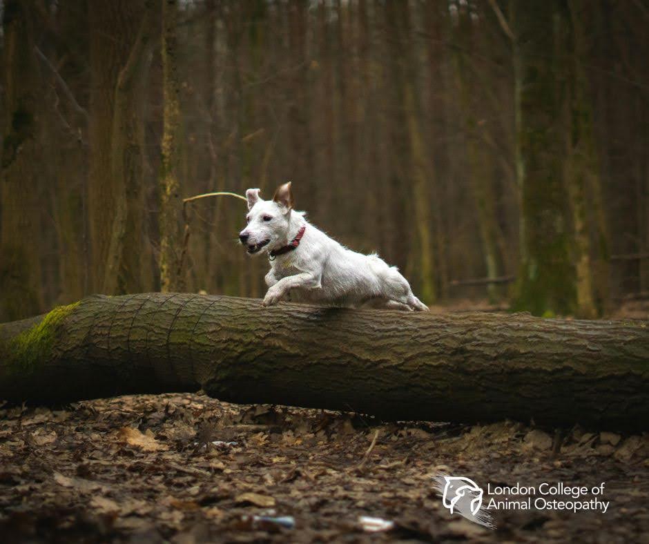
Walking the dog or riding their horse, owners may find that the dysfunction is not enough to be obviously classified as a pain reaction, but simply an alteration from the normal. Dogs may only show signs of fascial disturbance in ranges of movement that are beyond the normal range of usual gait assessment.
This is because canine fascia has been shown in dissection and histology studies to be quite similar to human fascia in that it is looser and less dense than equine fascia (Ahmed et al, 2019).
This development is perhaps due to the wider range of movement that canines have and a greater degree of flexibility. Therefore, while the vet may not find anything by observing them walk, the owner might find that their dog has changed their usual sleeping position for example.
We often see dogs curl into a sleeping position that alters the spinal curves and mechanics of the body beyond their range for normal walking and trotting; this could show dysfunction only when they attempt those positions.
The potential for fascia to be a factor in a more progressive dysfunction is clearly high, as it may not be spotted early and could progress to wider effects throughout the body before being given the attention it needs.
There is also the possibility that fascia becomes a maintaining factor in a pre-existing condition or injury. Fascia may be a part of the dysfunction, but another important factor is that it could be our “way in” when treating as well.
Osteopathy and Fascia
Osteopathy has always been ahead of the curve when considering a more open-minded approach and holistic view of healthcare. Fascia had been previously seen as a rather insignificant tissue by many professionals and considered to be inconsequential to injury or pathology.
However, as many Osteopaths already knew, the devil is in the details; it is often the most seemingly minuscule of observable disruptions that can create the perfect storm for larger, more “loud” symptoms. The old view of fascia was simply that of ignorance of the actual functions it has.
Models of Osteopathic intervention, such as the Cranial Osteopathy of W.G. Sutherland and techniques like balanced ligamentous tension (BLT), were acting upon the fascia in positive ways before the tissue was fully understood (not that it is fully understood yet).
The methods of balancing tensions through the matrix of interwoven collagen fibers and facilitating the perfusion and hydration of fascia could provide a multitude of positive outcomes if the fascia form part of the configuration of dysfunction (which of course it will as the body functions as a unit). It could be said that Osteopathy was ahead of its time in the understanding and treatment of fascial disruption.
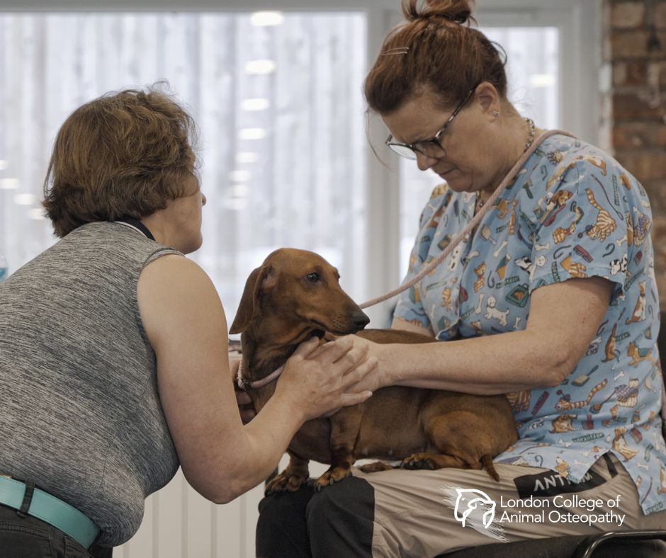
The well-honed skill of palpation, which is a hallmark of Osteopathy, gives a vast amount of information regarding fascia. When other approaches miss this aspect of assessment (or at least miss the depth that Osteopathy teaches), they can lose that vital data that informs their planning.
It can be easy to observe only the big movements and ranges when assessing, this is why Osteopathy trains the eye to seek the smallest of imbalance or asymmetry. This, combined with the Osteopath’s ability to gather a full and holistic history of the animal that considers all aspects of their life, Osteopaths have a great chance of finding the fascial disruption that could have been skipped over otherwise.
In Practice
In human studies (of which there are far more than animal ones), it is seen that lesions of the fascial tissue are highly prevalent in cases of muscular injuries associated with sports (Wilke, Hespanhol, and Behrens, 2019). Sports injuries are really just injuries related to either overexertion or repetitive actions, and these are things that animals will also be at risk of. It would be sensible to consider that fascial injuries would be equally prevalent in animal muscular injury.
Clearly the lines of distortion will be different in the animal models due to quadrupedal morphology. Some interesting models of fascial study have been created for animals to gain a better understanding of the forces transmitted through the tissues and how they may become damaged.
Through the dissection of animal cadavers, animal scientists have discovered that there are continuous connections throughout the body that are similar to those found in humans (The Fascia Guide, 2016). These “lines” as they are described are connections far reaching around the body and create a clear and measurable link between parts of the body that may have seemed too remote to have affected each other.
In horses, lines like the Dorsal line, which spans from the distal phalanx of the hind limbs, through the hamstrings, and attaches along the back to behind the jaw, demonstrate the interconnectedness of structures that owners might perceive as functionally separate. In Osteopathy, however, we use this principle of unity already, and our hypothesis is made upon a whole body assessment that will consider these connections.
In practice it is important to remember that these models are exactly that, “Models”. There is no such thing as a text book perfect animal and the unique variations of each individual are what need to be seen in detail.
If it were as easy as looking at the diagrams of fascial lines and deciding symptom “X” equals diagnosis “Y” then anyone with a text book could do it. The reality in practice is that cases will come to us with other practitioners having failed to resolve the issues because of having stuck to models and not principles.
Why the Principles are Important
The holistic nature of Osteopathy encourages us to see the wider picture. When we are assessing our patients, we can use tools such as the “Osteopathic Sieve,” which allows us to narrow down the presentation to certain tissues and then use that as a road map to find out how those tissues can’t cope with the load upon them.
We can “sieve” the nature of the presentation from its character of pain, biomechanical compensations, time scales of dysfunction, etc. This, combined with the knowledge of the structure and interrelated function of all the tissues and viscera, gives us all the clues to find the tissues causing symptoms.
We can then ascertain to what degree the fascia is impacted. Looking deeply enough, we could theoretically say that the fascia will always be affected to some degree when we consider the holistic functional unit of the body that Osteopathic principles teach.
The previous understanding of fascial lines and models is still very useful as we can visualise the state that one may expect in the fascia and then using clinical assessment we can compare that with what we actually find. This comparison can provide a way to measure the potential level of distortion however we shouldn’t be aiming for a visualised perfect, only a balanced and functional pain free “normal”.
From our Osteopathic philosophy, we can see that the state the fascia is in is the state of the body and not just the fascia. This allows us to create a treatment plan that can treat the entire patient and not just aim to “break down” adhesions or restrictions in fascia.
After all, the adhesions and restrictions are entirely correct for the configuration we have presenting in front of us; if we then deliberately remove those adhesions directly, the body will simply repeat itself or find another potentially more dysfunctional configuration.
With our philosophy guiding our treatment, the approach can be less aggressive on individual tissues, and continual palpation of change leads our technique to avoid unnecessary forces.
Technique in Action
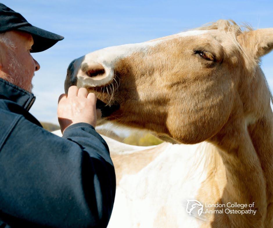
I personally have an experience of treating a horse that perfectly captures the nature of fascia and its importance in Osteopathic intervention. The horse in question was a 16-year-old TB X who had been displaying reduced performance in her flatwork undersaddle.
She had been seen by the vet who had been unable to find a specific cause and was suggesting that this could simply be riding related or overuse strain. The vet did not prescribe any medication or suggest any imaging and instead had referred to an Animal Physiotherapist and the Trainer to work together.
The Physiotherapist could not come up with a hypothesis for the riding problems, and the trainer was convinced that it was behavioural problems. When I was contacted by the owner, they were understandably fatigued by the ongoing problem without any ideas as to why.
I came to see the horse with veterinary permission and full cooperation from the Physiotherapist also. My first meeting with the horse was interesting, while there was clear concern from the owner during my case history taking, there was very little found on initial dynamic assessment so I asked to see her riding as this was when the issues were apparent.
Once the owner mounted, there was an instant change in the horse’s posture and behaviour. While many riding concerns show in a certain pace or with a particular exercise, this was clear from the moment the owner’s weight hit the saddle.
The horse instantly shifted her weight to her left. This can happen when mounting as normal from the near side, but the weight remained there rather than simply adjusting from the mounting. Observing the horse from the front, it was apparent that the head and neck had rotated on the frontal/coronal plan by about 5-10 degrees.
As the horse moved off into walk, I could see that the right bend was restricted (possibly due to the coupled motion it would produce), and there was a tendency to hollow the back and neck when making transitions up or down. Protraction of the forelimb appeared weak and lacked length in the cranial phase of all four limbs.
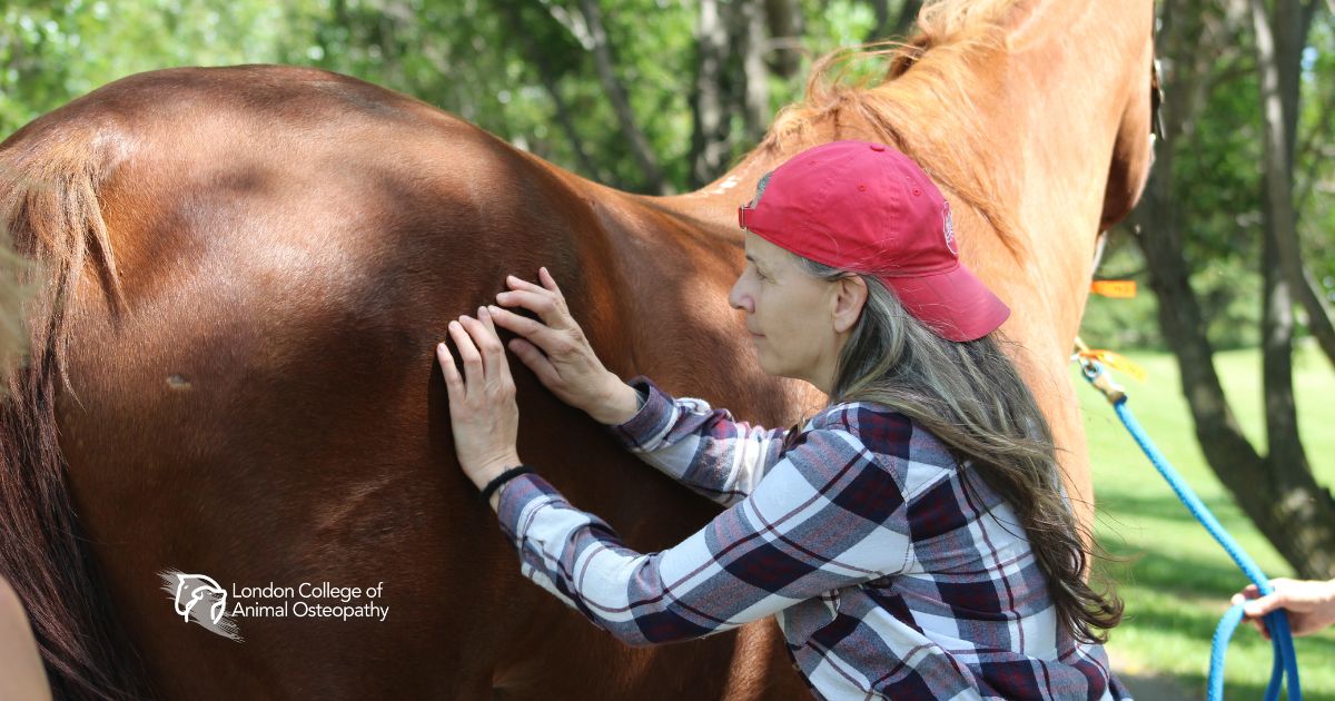
It was palpation that really informed me what was occurring. On returning to the stable to continue assessment, I palpated at the sternum and withers. I could perceive a sensation of fixation through the fascial diaphragm of the suprapleural membrane.
This dense and fibrous fascial layer separates the neck and thoracic cavity, supporting the regulation of cavity pressures. It could be visualised as the “lid” at the top of the ribcage. In Osteopathic palpation, we can develop the skill to detect the vectors of the distortions, including twist, stretch, compression etc. I could feel a left-sided unilateral distortion of twist and adhesion of the fibres in the suprapleural fascia.
I hypothesised that this was creating a left draw on the lower cervical spine, and when the weight of the rider was placed on top, the cavity pressure changes and exacerbates the pattern.
My treatment was very gentle, and using a form of BLT, I used compression of the tissues between my two hands to find the point of least resistance in that fascial layer. Once this point is achieved and held, there can then be a reorganisation of the tissues by the body itself.
This kind of technique can be learned within our masterclasses and courses at the London College of Animal Osteopathy. This treatment was profound in its effects but very gentle.
After the first treatment I suggested some stretching exercises and a limited riding regime that would allow for the pattern to remain unwound. After just three treatment sessions, the issues had fully resolved and the owner was over the moon.
It is clear that Fascia is equally important as the other tissues when assessing and treating. I could have tried articulating limbs and stretching muscles in a reductionist way, but the pattern creating those other restrictions seemed to stem from this fascial disruption.
Now, as this had been ongoing for a while and the owner couldn’t recall any trauma, we may never know what the initial cause of the pattern was. However, at this point (and in my own opinion) it almost doesn’t matter.
Osteopathy helps us to see the whole and the subtle; it trains the hands to perceive the most hidden of dysfunctions. Fascia is a fascinating tissue to study, and we still have yet to learn its full function. The goal is to remember that we are always students of the body, as A T Still said “Keep digging”.
Bibliography:
Ahmed, W., Kulikowska, M., Ahlmann, T., Berg, L.C., Harrison, A.P. and Elbrønd, V.S. (2019). A comparative multi‐site and whole‐body assessment of fascia in the horse and dog: a detailed histological investigation. Journal of Anatomy, 235(6), pp.1065–1077. doi:https://doi.org/10.1111/joa.13064.
Gatt, A., Agarwal, S. and Zito, P.M. (2020). Anatomy, Fascia Layers. [online] PubMed. Available at: https://www.ncbi.nlm.nih.gov/books/NBK526038/.
John Hopkins Medicine (n.d.). Muscle Pain: It May Actually Be Your Fascia. [online] www.hopkinsmedicine.org. Available at: https://www.hopkinsmedicine.org/health/wellness-and-prevention/muscle-pain-it-may-actually-be-your-fascia.
Starke, S.D. and Oosterlinck, M. (2018). Reliability of equine visual lameness classification as a function of expertise, lameness severity and rater confidence. Veterinary Record, 184(2), pp.63–63. doi:https://doi.org/10.1136/vr.105058.
The Fascia Guide. (2016). Fascia in Horses – Danish veterinary exploring uncharted territory. [online] Available at: https://fasciaguide.com/research/fascia-in-horses/.
Turner, S. (2024). Balanced Ligamentous Tension in Osteopathic Practice. Jessica Kingsley Publishers.
Wilke, J., Hespanhol, L. and Behrens, M. (2019). Is It All About the Fascia? A Systematic Review and Meta-analysis of the Prevalence of Extramuscular Connective Tissue Lesions in Muscle Strain Injury. Orthopaedic Journal of Sports Medicine, 7(12), p.232596711988850. doi:https://doi.org/10.1177/2325967119888500.