The Main Anatomy Of The Horse’s Back You Need To Know
The Main Anatomy Of The Horse’s Back You Need To Know
By Siun Griffin, Equine Physiotherapist and LCAO Community Manager
The anatomy and biomechanics of the horse’s back are essential for understanding how the horse moves and performs.
Having knowledge of the horse’s back structure is invaluable for horse owners and therapists alike. Ensuring that the back is kept in good condition with correct work, saddle fit, osteopathy, and vet care will help enable the horse to perform to its best whatever that task.
With this in mind, it is important to look at the whole horse as the back can affect other parts of the horse.
The back of the horse is a complex structure consisting of bones, muscles, tendons, and ligaments that work together to provide strength, stability, and flexibility.
Let’s take a closer look at its individual parts.
Lumbar Spine
The lumbar spine consists of six vertebrae that are behind the thoracic vertebrae and have no rib attachments. It starts after the last rib and 18th thoracic vertebrae. Several muscle groups and ligaments attach to the lumbar vertebrae.
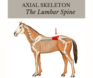
It has large transverse processes, and wide, flat vertebrae. This gives the lumbar area limited mobility. These connect to the much more mobile lumbosacral joint that provides hind-end propulsion. However, its latero-flexion mobility is limited.
The muscles that support the lumbar spine include the longissimus dorsi, iliocostalis, and psoas muscles.
The ligaments of the lumbar spine include the supraspinous and interspinous ligaments, which provide support and stability to the spine.
This tends to be a weaker area of the spine as it does not have the support of the ribs. This is one area where saddle fit is important as it should never sit on the lumbar spine.
Sacral Vertebrae
After the lumbosacral joint, you have the sacral vertebrae which slope slightly towards the tail. This section of the spine has fused vertebrae and doesn’t flex at all. It acts as one bone. Here, major ligaments and muscles attach from the pelvis and this section helps to support the hindlegs.
After the sacral vertebrae, the last section of the spine is in the tail. Horses can have 15 to 25 of these small vertebrae in their tail, with an average of 18.
Thoracic Spine
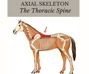
The thoracic spine of the horse consists of 18 vertebrae (T1 – T18). The withers are part of the thoracic spine and are the largest spinous processes. It has limited mobility and is supported by large muscles, ligaments, and ribs.
The muscles that support the thoracic spine include the longissimus dorsi, multifidus, and serratus ventralis muscles.
Main Muscles of the Back
The main muscles of the horse’s back which span across multiple sections of the spine include the longissimus dorsi, multifidus, serratus ventralis, iliocostalis, spinalis thoracis, and trapezius.
Longissimus dorsi

The longissimus dorsi muscle supports the lumbar spine and runs from the sacrum to the withers. It can act to extend and laterally flex the spine and is the largest muscle of the horse’s back.
The longissimus muscles are the easiest to use to see and feel as they are found on the surface. Horses often get sore, develop spasms, and get fatigued in these muscles as they do so much to support the horse.
Multifidus muscle
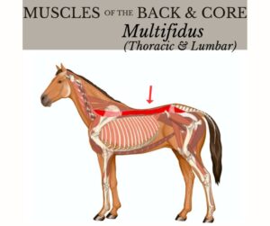
The multifidus muscle runs along the spine and provides support and stability to the vertebrae. This is a deeper muscle and divides into segments that support only a few vertebrae each.
It is important that the multifidus functions correctly and is strong, otherwise too much pressure is put on the longissimus to provide support. When that happens, you get the issues mentioned above in the longissimus.
Serratus ventralis

The serratus ventralis muscle is located on the sides of the spine and provides support and stability to the rib cage. It is a large, important muscle that supports the chest and breathing.
The dorsal section of this muscle sits close to the spine. The cranial part of the dorsal section is found behind the scapula and over the first few ribs. It sits under the latissimus dorsi, except for the lowest part, which is just under the skin. (The area where the rider’s leg sits).
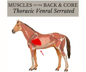
Both the thoracic and cervical sections of the serratus play a major role in leg and shoulder movement. It is important for the health of the back as poor saddle fit can affect it, thus having a knock-on effect on the entire back.
Trapezius
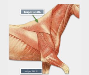
The trapezius has two sections, cervical and thoracic. The thoracic is a triangular shape that sits over the withers and attaches to the scapula. It is not a weight-bearing muscle, but the saddle does sit on the caudal portion. Poor-fitting saddles can cause a lot of damage to the trapezius if they put excess pressure on it.
Its main job is to move the shoulder blade.
Iliocostalis muscle
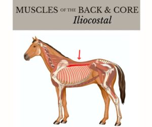
The iliocostalis muscles are located on the sides of the spine and provide support and stability to the back. It is divided into segments that run from the lumbar spine to the third rib.
When these muscles contract on both sides, they help extend the spine. When one side contracts, you get lateral bending.
On many horses, it is possible to palpate these muscles.
Spinalis dorsi muscle
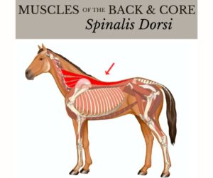
The spinalis dorsi muscle is a deep muscle. It is responsible for supporting the vertebrae and movement of the neck. It runs from the lumbar to the cervical spine.
It plays an important role in stabilizing the cervico-thoracic junction. It is underneath the trapezius and often doesn’t get much attention or thought.
Poor saddle fit can cause quite a bit of damage to this muscle. So it is important not to forget about it.
Iliopsoas muscles
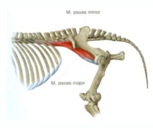
While not a main back muscle of the horse, this muscle group does help stabilize the spine as well as protract the hind limbs and helps with hip flexion. They are deep muscles, making them hard to treat and identify as the source of pain. They are vulnerable to strain. An effective technique for reaching the iliopsoas is the OAB technique.
Ligaments of the horse’s back
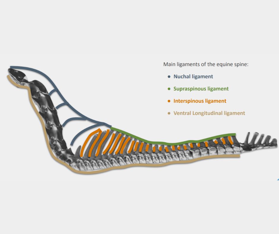
Interspinous ligament
This ligament runs along the spine filling the spaces between the spinous processes while helping to support the spine’s vertebrae in movements such as flexion and extension. It is this ligament that interspinous ligament desmotomy is performed for some horses with kissing spines.
Supraspinous ligament
The supraspinous ligament is a strong, important ligament that helps prevent the vertebrae from moving too much, and provides support. It is long, running from T3 to the sacrum.
It sits on top of the dorsal spinal processes and is a continuation of the nuchal ligament that runs from skull to the withers. This ligament does not have a lot of elasticity and can be injured when overstretched.
The widest section sits over the withers where many muscles attach to it.
Ventral longitudinal ligament
The job of this ligament is to prevent the spine from overextension. It sits below the vertebrae.
Basic Biomechanics
The horse’s back is essential for maintaining balance, stability, and flexibility during movement. When the horse moves, the muscles of the back work together to support the weight of the rider. The back also plays a vital role in the horse’s movement, allowing it to perform a range of movements, including collection, extension, and lateral movements.
The biomechanics of the horse’s back are complex and depend on the interplay between the bones, muscles, tendons, and ligaments.
The muscles of the back work together to provide strength, stability, and flexibility, while the tendons and ligaments provide support and stability to the spine. The shape and structure of the spine also play a role in the biomechanics of the horse’s back, allowing for a range of movement while maintaining balance.
The anatomy and biomechanics of the horse’s back are complex and essential for understanding how the horse moves and performs.
While this isn’t an in-depth look at the horse’s back it is a good starting point for anyone who works with horses to develop their understanding of how it works. Having this knowledge will help equestrians better manage their horses and help them stay comfortable.
Special thanks for graphics credit: EquiPro Connect
Gain an in-depth understanding of Equine Functional Anatomy & Biomechanics here