Animal Meridians: Energy Pathways & Fascial Therapy

By Chris Bates M.Ost, DipAO, EEBW, BHSAI

The term “meridians” is used across the therapeutic industry to describe a number of things. The interchangeable use can create confusion around what a therapist is actually doing and the models they are using to assess and treat an animal. The fact remains however that the word is very useful to describe both energetic phenomena and the anatomical pathways of certain tissues. It can be very useful as owners to understand more deeply what their therapist is referring to and this article aims to clarify what these terms and models mean.

Understanding the Body as an Integrated Whole

The body of any animal can be described from a variety of angles. Notice I didn’t say it can be “separated out” or “broken down”, this is because I’m an Osteopath and we know that separation and isolation of systems means it is no longer a “body”. The different angles one can view the body will inevitably lead to different models of health. We could think of models of health as our way into intervention; we have to start somewhere and hopefully gain a greater understanding holistically with more information.

What Does Holistic Really Mean?

The term “holistic” gets thrown around a lot in the world of health but rarely does it actually mean addressing the whole. We see myofascial therapists referring to myofascial release as holistic, but this could only be true if the entire body was muscle and fascia…

Of course this does not mean that those therapists do not think holistically, nor does it mean that the therapy is invalid. But we need to see modalities as “ways in”, not as independent. All therapies will affect the other aspects of the being because animals (and we) are integrated units, myofascial release will affect the mind, acupuncture will affect the connective tissues, it is unavoidable and of course essential.

Now that’s cleared up (well, as clear as mud for most of us), let’s discuss some of these “angles” and how they work.

The Energy Model Meridians

Traditional Chinese Medicine and Qi Flow

The concept of energy meridians can be traced back to early traditional Chinese medicine (TCM). This concept was transported around Asia and also forms the basis of the Japanese system of Shiatsu (finger pressure). This energy concept describes a universal energy and life force existing in all living beings called Qi (Chi – Chinese, Ki – Japanese).

The health of the individual depends on the optimal flow of this energy throughout the body via meridians which are described as vessels or pathways for this energy. If this flow becomes blocked, weak or even excessive, then physical and emotional effects can appear.

The 12 Meridians and Acupoints

In TCM there are 12 meridians that correlate to an organ or system plus additional meridians that have more central functions such as the “governing vessel” and “conception vessel” which run along the spine. Along these meridians are points at which one can intervene in cases of dysfunction, these are acupoints and practitioners will use needles (acupuncture) or pressure (acupressure/Shiatsu) to clear blockages and mediate flow.

Ayurvedic Nadis and Prana

There are actually many similarities in the theory of TCM meridians and the traditional Indian system of Ayurveda. In Ayurveda, the pathways of this energy are called Nadis and the energy flowing throughout is referred to as Prana. There are similarities in Ayurvedic treatments and TCM/Shiatsu however there is often more emphasis within Ayurveda on the internal systems of control such as breathing techniques (Pranayama), nutrition and how one conducts themselves.

Obviously teaching an animal a breathing technique is rather difficult, Ayurvedic principles have however been applied to the animal model and specific modalities created. It could be argued that one could observe dysfunctional breathing patterns in an animal who is experiencing energetic dysfunction though. We must remember that a model is not just for intervention techniques but how we assess and examine, an “angle” to see from.

The Connection Between Energy Pathways and Anatomy

Although the principles of energy meridians are different to the biomechanical models, some of the energy pathways do seem to follow the path of major blood and lymphatic vessels or nerves. Some meridians being closely correlated with neurovascular bundles showing there could be a connection to their function.

As discussed, the body cannot be broken down and separated or it is no longer a body, so it comes as no surprise that this is the case because one approach will influence other systems. If we widen our lens viewing the energy meridians, we see that there is also some correlation with connective tissue pathways such as fascial lines. This is not an attempt to “explain away” the energetic principles as phenomena of the anatomy, but rather a way to highlight the validity of the approach.

Why Energy Meridians Work Well for Animals

When we look at how the intervention at meridians is aimed we can see the usefulness in animal practice clearly. Acupressure and acupuncture both deal with meridians but trying to address the entire system by the effects on just the energy system.

In TCM and Shiatsu, imbalance and poor flow of energy can lead to behavioural changes and emotional issues, with animals this can be particularly useful as an approach as unlike humans, they cannot receive talking therapies or coaching to overcome these states. Again, we have a “way in”.

Animals are particularly sensitive to touch and movement being that these are communication tools of the highest order in their evolution. The somewhat more subtle and less assertive methods of energy intervention can be a more suitable way to deal with animals who do not tolerate harder touch or too much contact.

Fascial Lines

What are Fascial Meridians?

Sometimes described as fascial meridians, the lines that appear in the fascial connective tissue in animals play a different role to the energy lines. The fascia is a collagen rich connective tissue which wraps and binds pretty much everything else in the body.

It plays a number of roles such as support, communication of forces, fluid transfer and it is even piezoelectric contributing to signaling. Our quadruped animals need to distribute their weight differently to ourselves as bipeds. This is why although there are many similarities with human fascia, animals rely on different fascial meridian layouts.

Understanding Fascia in Practice

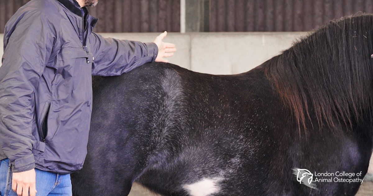
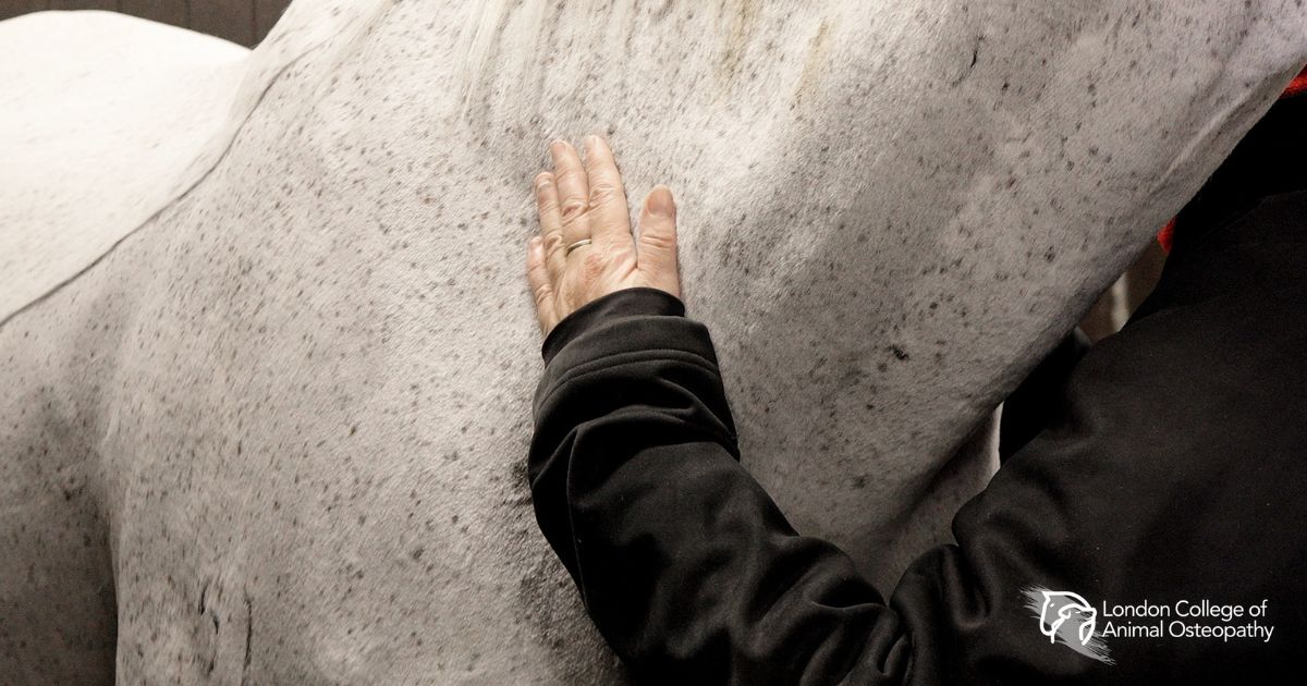
Many will have seen fascia if you have bought meat from the butcher and seen the silvery/white covering of connective tissue and you will have felt it if you have placed your hand on a horse’s twitching shoulder. Although there seemed within the therapist community to be a push towards making out that fascia was some kind of newly discovered “holy grail” in healing, the reality is that this is not new and fascia has always been impacted by the approaches of manual and physical therapists.

Of course our understanding of fascia has improved with research and this serves to allow us a clearer understanding around why our approaches work so well.

Key Fascial Lines in Animals

If one were to look at a map of fascial lines annotated onto a picture of a horse or dog, they would not look too dissimilar to some of the energy diagrams. But this model uses tracking of fascial forces and tensions. This is again just another “way in” to the whole.

The Superficial Dorsal Line (SDL)

One fascial pathway often referred to within manual therapy is the superficial dorsal line or SDL. This line tracks from the occiput (caudal skull), along the dorsal aspect, down the caudal aspect of the hind limb and into the plantar fascia. The SDL aids in the stored force required for propulsion and elasticity in that movement and also supports posture, blending with very strong structures like the nuchal ligament and supraspinous ligament.

Dysfunction in this SDL can lead to lack of hind end engagement, reluctance in spinal extension and postural impacts.

Frontal and Spiral Lines

Conversely there are frontal or core lines and the superficial ventral line that support truncal flexion, breathing, digestive function and assist in guarding and anxiety related behaviours. The spiral lines assist in torque required for rotational movement and complex body movement coordination, dysfunction here can show in lateral asymmetries (static and dynamic) and difficulty in tight turns.

Signs of Fascial Disruption

It’s clear to see there are a diverse range of signs and symptoms that can be observed in fascial disruption. When one considers that fascia not only follows lines of force through the musculoskeletal system but also interconnects with the viscera, vessels and nerves, it becomes apparent to us that there are far reaching systemic effects of fascial disruption too.

How is Fascia Treated?

So how is it treated? Fascia responds well to light sustained touch when tethering or restriction are present. Slower movements and gliding touch can encourage layers of the fascia to slide and alter the friction coefficient by influencing the distribution of hyaluronic acid (the lubricating gel-like substance found within fascial tissue).

Therapists also claim that myofascial strokes improve hydration of the fascia in general but this claim is less researched; Osteopathically it does make sense as improving motility and vascular perfusion should allow for fluid transfer changes. These slower movements and softer touch do seem to have positive influences however, are these purely due to fascial “unwinding” and stretching? Osteopathy would argue, No…

The Osteopathic Perspective of Both Meridian Models

Using Different “Ways In” to See the Whole

As is repeated throughout this article, we are using “ways in” to the whole. When eating a meal, we don’t just engulf the whole thing in one go or we choke. We take smaller bites until we have the entire thing digested. This is how the picture of an animal’s health appears to us.

In Osteopathy we want to obtain a picture of the whole animal and their complaint. Do we look at the biodynamics or the behaviours? Do we look at the posture or the pain? The answer of course is that we need to look at all of those but we need to metaphorically “move around” to see all those angles.

The Osteopathic Assessment Approach

Within that “moving around” we will use assessment tools such as fascial lines, dynamic assessments and indeed we may look at energy meridians (if we have training in that area). Osteopathy is not so much about trying to eat the metaphorical meal in one bite, it’s about understanding there is a whole meal. By understanding the unity of all the systems and structures, we know that our hypothesis will only be a guide to what is actually happening.

Benefits of the Energy Model in Osteopathy

Energy work and Qi/Prana flow are an intriguing way to see the body and there are some benefits to observing from this traditional medicine approach. One plus to this model is that a meridian that is located in one part of the body will influence systems away from that location. Rather than simply accepting that as the way it is, in Osteopathy we can ask “why?” and investigate the interconnectedness through all the potential pathways.

Another plus is that energy work seeks to remove blockages and barriers to flow, this is exactly what Osteopathy does too. Rather than putting in something seen as missing or extracting an illness as if it were an entity on its own, Energy work promotes the animal’s own intrinsic healing mechanisms.

How Fascial Lines Inform Osteopathic Practice

Fascial lines/meridians show us as Osteopaths another tissue that could be affected by trauma or systemic imbalance/allostasis. Osteopathy has always been about deeply understanding anatomy. The better our knowledge of anatomy, the more we can hypothesise where health may be hindered.

Fascia feeds perfectly into our concept of bio-tensegrity, it offers Osteopaths another avenue beyond muscle connection, tendon and ligament to visualise forces being transmitted throughout the body.

Explaining Connections to Owners and Vets

Osteopathy used the term somatic dysfunction and models such as somatovisceral and viscerosomatic reflexes to explain certain presentations; Fascia offers an explanation for the connections that may (without it) seem tenuous and difficult to rationalise with owners and Vets.

Osteopathy remains open to new discoveries and to the traditional approaches alike. This is a strength in practice by not becoming biased to one model.

Supporting Elite Dressage Horses with Osteopathy

23 Jan 2026 Equine

Click to view


Elite dressage places extraordinary demands on the horse. Pirouettes, piaffe, passage, tempi changes — each of these movements loads the hindlimbs, strains the lumbar-sacral region, and requires sustained cervical flexion that, over time, can tip from healthy exertion into chronic dysfunction. Dr. Amelie Villeneuve-Moore’s rigorously researched thesis asks a precise and important question: Does equine osteopathy have a specific role to play in supporting dressage horses, and if so, what does that look like in practice?


Through an extensive literature review of 44 papers, Dr. Villeneuve-Moore identifies two dominant patterns of dysfunction in elite dressage horses: hindlimb suspensory ligament injuries, which can be career-ending, and myofascial trigger points — particularly in the cervical and thoracic regions — which can silently limit performance without causing outright lameness. Dressage horses, the research reveals, carry significantly more myofascial trigger points than showjumpers, with the brachiocephalic and cervical muscles most commonly affected. These findings have direct implications for how osteopaths assess and treat top-level performance horses.


This thesis presents a compelling argument that routine osteopathic interventions can significantly reduce injury risk and aid in the rehabilitation of horses. It assesses various osteopathic methods, including ischemic compression, longitudinal myofascial stretches, and OAB-based systemic rebalancing. This work is highly relevant and sophisticated, offering valuable insights for equine osteopaths, veterinarians, and dressage professionals.

Comparative Osteopathy for Canine Dysfunction

5 Dec 2025 Canine

Click to view


Some of the most clinically challenging cases in veterinary practice are those where something is clearly wrong — a dog moves differently, holds itself awkwardly, reacts to touch in ways that suggest discomfort — but where imaging produces nothing conclusive. These are the patients that idiopathic somatic dysfunction describes: functional impairment without identifiable structural pathology. Jayne Strange’s thesis asks a pointed question: can osteopathic treatment, informed by the richer evidence bases of human and equine medicine, be effectively adapted to help these animals?


The comparative framework Strange employs is rigorous. She maps somatic dysfunction across three species — humans, equines, and canines — identifying shared mechanisms of myofascial tension, segmental restriction, proprioceptive disturbance, and neuromuscular imbalance. The fascia emerges as a common thread: a mechanosensitive tissue that responds to manual input in ways that are now well-documented in humans and increasingly observed in horses, and which almost certainly operates similarly in dogs.


The equine evidence base receives detailed attention. Research by Haussler, Clayton, and Stubbs demonstrates that spinal manipulation in horses improves stride length, reduces muscle asymmetry, and enhances thoracolumbar mobility — outcomes measured through kinematic analysis rather than subjective observation. Strange draws a careful line from these findings toward the canine, noting that while the species differ in scale and loading patterns, the longissimus and multifidus muscles play comparable stabilising roles in both, and that compensatory movement patterns following restriction look strikingly similar.


Human clinical trials — including a landmark meta-analysis of nearly 900 patients with chronic low back pain — provide the mechanistic grounding that veterinary research cannot yet offer for dogs. Techniques such as myofascial release, muscle energy technique, and balanced ligamentous tension have demonstrably modulated pain, improved proprioceptive feedback, and restored functional mobility in humans. Strange makes a reasoned case that these mechanisms are biologically transferable, even if their dosage and application require species-specific adaptation.


The thesis is clear-eyed about what the canine literature currently lacks — standardised diagnostic protocols, objective outcome measures, and controlled clinical trials. But Strange frames this as a call to action rather than a limitation. The question she leaves open — and which practitioners will find genuinely useful — is how to build that evidence base while responsibly treating the patients in front of us now.

Osteopathic Insights into Post-Castration Complications

17 Nov 2025 Equine

Click to view


Castration is the most commonly performed surgical procedure in equine practice. It is also one of the most routinely dismissed when it comes to long-term consequences. The scar heals, the horse goes back to work, and the surgical site is rarely examined again. Yet equine osteopaths have long observed that geldings sometimes present with unexplained movement asymmetries, hindlimb stiffness, and pelvic dysfunction that conventional diagnostics cannot explain — and that often improve when the castration site is addressed. Pauline Bürgermeister decided to put that clinical intuition to the test.


This thesis presents an original study of 25 geldings — a mix of Standardbred trotters and riding school horses — in which castration scars were systematically assessed for five clinical signs: depression of the scar tissue, fibrotic scarring, pain on palpation, discharge, and oedema. Gait asymmetry was evaluated using Sleip AI, a validated markerless motion analysis system, capturing head, pelvic, and total asymmetry scores. The results are striking.


The composite clinical score showed a strong positive correlation with pelvic asymmetry (ρ = 0.71, p < 0.001) and a moderate correlation with total asymmetry (ρ = 0.53, p = 0.007). Tissue depression, fibrotic scarring, and oedema were the most influential individual signs. Pain alone did not reach significance — suggesting that the most clinically relevant changes are structural and chronic rather than acutely painful. Discharge appeared to dilute the composite score, highlighting the importance of weighting variables appropriately in future work.


The thesis contextualises these findings within osteopathic anatomy, examining how fascial restriction, impaired lymphatic drainage, and the well-documented formation of neuromas in spermatic cord remnants might mechanically and neurologically contribute to pelvic dysfunction. The study is honest about its limitations — small sample, no blinded assessment, heterogeneous population — and frames its findings as a call to action rather than a conclusion. For any practitioner encountering unexplained hindlimb asymmetry in a gelding, this work offers a compelling reason to look further down the leg.

How does Osteopathic Manual Therapy (OMT) support and improve sport horse performance?

13 Oct 2025 Equine

Click to view


The demands placed on an elite sport horse are extraordinary. Whether jumping combinations at speed, performing collected dressage movements, or racing under load, these animals are required to operate at the edge of their physical capacity — often day after day, season after season. It is no surprise that managing their health requires more than a single discipline. Josephine Jansen’s thesis makes a careful and well-supported argument for why Osteopathic Manual Therapy should occupy a central role in that broader care picture.


Jansen opens with a clear articulation of what osteopathic treatment actually means in the equine context — not symptom management, but whole-body assessment aimed at identifying the primary cause of dysfunction and restoring homeostasis. She draws on Still’s founding principles to frame OAB (Osteopathic Articular Balancing) not as a technique in isolation, but as a neurophysiological intervention: one that alters signals within the central nervous system, influences sensory processing, and can interrupt chronic pain cycles that persist long after the clinical signs of injury have resolved. The concept of ‘traumatic muscle memory’ — neural activity that continues reverberating in the CNS even when tissue has healed — is introduced with precision and practical relevance.


Two significant studies form the evidentiary backbone of Jansen’s argument. A French study of 26 sport horses found measurable improvements in propulsion, symmetry, and regularity of gait following a single osteopathic treatment, with younger horses showing the most immediate response and older horses benefiting from repeated sessions. A larger study by Thoresen, examining 374 horses with suspected sacroiliac, hip, and back dysfunction, found positive outcomes in approximately 80% of cases — including horses that had not responded to intra-articular injections.


The final section on integrative medicine is where the thesis becomes most forward-looking. Jansen draws on research from equine sports medicine to argue that the best outcomes for performance horses emerge not from any single modality but from a coordinated team — veterinarian, osteopath, farrier — each contributing their specialist lens to the same problem. The hoof’s relationship to upper musculoskeletal health, the limitations of traditional diagnostics in subclinical cases, the role of preventive care in extending a horse’s competitive career: all are addressed with clarity and practical grounding.


This is a thoughtful, well-researched thesis with a strong clinical voice. It neither overclaims nor undersells, and for practitioners working in or adjacent to performance horse care, it provides both the evidence and the rationale to advocate for OMT as a serious, integrated tool in sport horse practice.

Osteopathic Manipulative Therapy in the Management of Trigeminal Mediated Headshaking in Horses.

29 Sep 2025 Equine

Click to view


For horses affected by trigeminal-mediated headshaking, the prognosis is sobering. The condition — characterised by violent, involuntary vertical flicking of the head, nasal irritation, and profound distress, often triggered by exercise, light, or wind — has no confirmed cause and no reliably curative treatment. When EquiPENS, supplementation, and pharmacological options fail to bring meaningful relief, euthanasia is frequently the outcome.


Sadie Pearce’s thesis does not offer a clinical trial or a treatment protocol. What it offers is something arguably more valuable at this stage: a carefully constructed hypothesis. Through a systematic narrative review of the available literature, she maps the anatomy and neurophysiology associated with TMHS and asks whether the physiological changes described in osteopathic somatic dysfunction could illuminate — and potentially address — the underlying pathology.


The trigeminal nerve is the largest cranial nerve, with extensive sensory coverage across the head. Research has confirmed hyperexcitability of its maxillary branch in affected horses, with measurable reductions in the threshold at which the infraorbital nerve fires. What has not been established is why. Pearce traces the anatomical connections between the occipitoatlantal joint, the superior cervical ganglia, and the trigeminal nerve’s origin in the brainstem — a pathway where osteopathic lesioning, she argues, could plausibly contribute to the dysfunction observed.


The review draws on spinal lesion research, fascial mechanics, neurological signalling, and even the emerging evidence around gut microbiota differences in TMHS horses, building a layered picture of a condition that may be systemic in origin rather than localised to the nerve itself. The role of calcium and magnesium in neural excitability, and the known therapeutic effect of magnesium supplementation, adds further texture to the hypothesis.


Whether OMT could offer these horses a meaningful pathway to relief remains an open question — but this thesis makes a compelling case that it deserves serious investigation. The full paper is worth reading for anyone working with horses for whom current treatment options have run out.

Osteopathy in Foals and Growing Horses

30 Jun 2025 Equine

Another standout work by an LCAO student! Read about Osteopathy and its effects on foals and growing horses in a thesis by International Diploma in Equine Osteopathy student, Claire Delisle Legrand.


The Use of Neurological Segmentation in Equine Osteopathy

7 Apr 2025 Equine

Click to view


Touch a vertebra, and you are not just touching a bone. You are entering a neurological network that extends outward to skin, organs, muscles, and the brain itself. This is the central insight of Frouke-tea Friedhoff’s thesis, which argues that equine osteopaths who understand neurological segmentation can address the underlying cause of dysfunction rather than managing only its visible symptoms.


The paper moves methodically through the architecture of the equine nervous system — central and peripheral, motor and sensory, somatic and autonomic — before arriving at the heart of its subject: the ways in which spinal segments, somatotopic maps, referred pain patterns, and dermatome regions can each guide more precise osteopathic diagnosis and treatment.


Particularly compelling is the discussion of spinal segmentation in practice. Working on the thoracic region at Th1–Th3, for example, carries implications not only for the vertebrae themselves but for the lungs, the heart, and the brachial plexus. The practitioner who knows these connections treats with intention rather than by location alone.


The thesis also explores somatotopic mapping — the idea that each region of the body corresponds to a specific point in the nervous system and in the cortex. Drawing on both human research and emerging equine-specific auricular acupuncture maps, Friedhoff suggests these tools could significantly enhance osteopathic diagnosis, helping distinguish whether a spinal lesion originates from the vertebral column or from organ dysfunction.


Referred pain in the horse is examined with appropriate caution — it is difficult to detect and often overlooked — but the author makes a persuasive case that ignoring it risks misattributing lameness to the wrong source. A dull coat, an unusual hair pattern, a subtle asymmetry: Friedhoff shows us how to read these as neurological signals. The thesis ultimately invites practitioners to look past the surface and ask: what is the body actually telling us?

1 2 3 4

Search

+